Moderate to severe hypercalcemia associated with hyperthyroidism is not very common and can raise a diagnostic challenge to the treating physician.
To present a case of thyrotoxicosis manifested with symptoms of hypercalcemia and resolution of symptoms after rendering euthyroid.
We report the case of a 61 year old Omani gentleman, who presented with symptoms of hypercalcemia like weight loss (10 kg) with poor appetite, constipation and depression as first manifestation of hyperthyroidism.
His lab investigations revealed moderate hypercalcemia ,corrected calcium was 3.04 mmol (normal 2.1 to 2.6 mmol) PO4 1.31 mmol (0.7 to 1.5) with suppressed parathyroid hormone level (PTH) 0.7 pmol (1.6 to 6.9 pmol). Thyroid function test (TFT) showed-TSH 0.003 mIU/l, FT4 70 pmol (8.5 to 22.5 pmol). Neck US showed diffusely enlarged thyroid with two hyperechoic nodules 2 × 1.3 and 2.2 × 1.5 cm and thyroid scintigraphy showed (99 mTc uptake) increased uptake 14% with multiple hot nodules. Diagnosis of hypercalcemia secondary to toxic multinodular goiter was made. He received saline hydration, palmidronate infusion and anti-thyroid therapy-(carbimazole). His calcium level dropped to 2.4 mmol on 5th day of admission.
Hyperthyroidism can be presented with symptoms of hypercalcemia. Clinicians should be aware of this unusual association, so that a prompt initial evaluation and proper intervention can be administered.
Hypercalcemia, Thyrotoxicosis, Ttoxic multinodular goiter, PTH.
Primary hyperparathyroidism and malignancy are by far the most common causes of hypercalcemia in our clinical practice [1]. Hyperthyroidism can usually cause minimal elevation of serum calcium. Asymptomatic serum calcium elevation has been documented in up to 20% of patients with hyperthyroidism and is related to increased bone resorption [2] and subsequent release of calcium from the bone into the circulation. We describe a case of hyperthyroidism presented with symptoms of hypercalcemia.
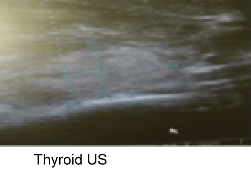
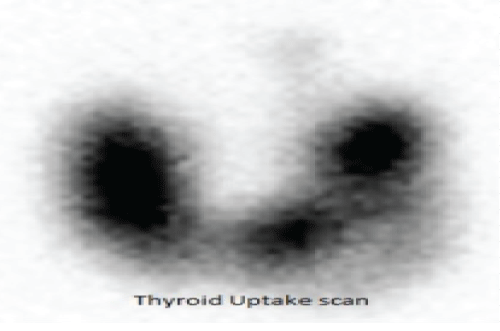
A 61 year old Omani gentle man, chronic smoker with past history of peripheral vascular disease, presented to our emergency department with 2 month history of weight loss (10 kg) with poor appetite, constipation and depression. There was no history of fever, hemoptysis, palpitation, and tremor or heat intolerance. Initially he was admitted under internal medicine team. Clinically he was thin built with no pallor, clubbing or lymphadenopathy. Pulse rate was 78/minute and regular; BP was 132/78 mm of Hg. He had small palpable goiter with no thyroid associated orbitopathy or dermopathy. Systemic examination was unremarkable. His lab investigations revealed normal complete blood count, ESR, C-reactive protein, glucose, renal, liver function and 25 -OH vitamin D level. Corrected calcium level was 3.04 mmol (2.1-2.6 mmol) with PO4 1.31 mmol (0.7-1.5), alkaline phosphate 183iU/L (40-150) and parathyroid hormone level (PTH) 0.7 pmol.(1.6-6.9). Thyroid function test (TFT) showed-TSH 0.003 mIU/l, FT4 70 and FT3 33 pmol. He was transferred to endocrine team. Neck US showed diffusely enlarged thyroid with two hyperechoic nodules measuring 2 × 1.3 and 2.2 × 1.5 cm in both lobes (Figure 1) and thyroids cintigraphy showed (99 mTc uptake) increased uptake 14% with multiple hot nodules (Figure 2). The investigations to rule out the possibility of malignancy including CT neck chest and abdomen showed normal study.
 Figure 1: Hyper echoic nodule 2 × 1.3 cm.
View Figure 1
 Figure 2: Thyroid uptake 14% with multiple hot nodules.
View Figure 2
Diagnosis of hypercalcemia secondary to toxic multinodular goiter was made. He received intravenous saline, palmidronate infusion (bisphosphonate), and carbimazole 15 mg three times per day with daily serum calcium monitoring. His calcium level dropped to 2.4 mmol on 5th day of admission. On follow up after 8 week she gained 3 kg, and was euthyroid clinically withnormal TFT and calcium. He was referred to Nuclear medicine team for radioactive iodine ablation in view of toxic multinodular goiter. Fine needle aspiration biopsy (FNAB) taken from photopenic area showed benign cytology (Thy2). As nodules were hot no biopsy taken from the nodules.
Primary hyperparathyroidism is the most common cause of asymptomatic hypercalcemia and is seen in ambulatory patients, whereas hypercalcemia secondary to malignancy is seen in hospitalized patients and usually symptomatic. Hypercalcemia in thyrotoxicosis is typically mildwith calcium levels rarely exceeding 4 mmol [3]. Our case demonstrates hypercalcemia merely due to thyrotoxicosis, as supported by low parathyroid hormone (PTH) level and resolution of hypercalcemia with treatment of the thyroid disease.
Our literature review revealed multiple case reports of concurrent primary hyperparathyroidism in association with hyperthyroidism. Albert Shiehand Dorothy Santos Martinez reported a similar case in endocrine society's annual meeting [4]. Korytnaya E and colleagues reported a similar case with vitamin Deficiency [5]. However, our patient's presentation was unusual as apathic without any adrenergic symptoms. Only one case report in the literature showed presentation with symptoms of hypercalcemia [6]. A linear correlation seen between serum calcium levels and levels of thyroidhormone, which is more pronounced in people over 60 years [7]. Our patient was 61 year old and had moderate hypercalcemia (3.04) relative to thyroid hormone level. As hyperthyroidism is associated with increased bone turnover, high levels of serum alkaline phosphatase (ALP) mostly of bone origin are usually detected in thyrotoxic patients [8]. Our patient had raised ALP of 183 iU/lL. Thyroid hormones are known to cause bone resorption and mobilizing calcium from bone to circulation leading to hypercalcemia. High levels of interleukin-6 (IL-6) seen in hyperthyroidism stimulates osteoclastic activity and alter osteoblast osteoclast coupling. Triiodothyronine (T3) known to increase the sensitivity to IL-6 and leads to accelerated bone resorption [9,10]. Thyrotoxicosis can be overlooked as in our patient if present with symptoms of hypercalcemia like constipation depression and loss of appetite.
Clinician should be aware of the association of hypercalcemia with hyperthyroidism. This will facilitate early diagnosis and appropriate intervention.