Autoimmune, Pancreatitis, Pseudotumoral
Immunoglobulin G4-autoimmune pancreatitis (IgG4-AIP) is a relatively rare chronic inflammatory disease of the pancreas. The physiopathology of this disease is poorly understood and still unclear. Due to the lack of specific clinical symptoms and imaging findings, it is often misdiagnosed as pancreatic cancer which may lead to abusive surgery.
Herein we are report a rare case of IgG4-AIP in a 70-year-old woman, admitted in hospital for chronic epigastric pain. Clinical investigations and CT-scan revealed a pancreatic-head mass, and scan guided biopsy was then assessed. Histological examination showed a fibrotic tissue rich in a dense infiltration with a large number of chronic infalmmatory cells. Mainly lymphocytes and plasma cells, with no histological signs of malignancy (Figure 1A). The immunohistochemistry staining showed the lack of CKAE1/AE3 as well as intense staining of plasma cells with CD68, CD138, IgG, and IgG4 antibodies (Figure 1B, Figure 1C, Figure 1D, Figure 1E and Figure 1F). Thus, final diagnosis of IgG4-AIP had been made and the patient was recovered after oral glucocorticoids treatment. This case highlights the unusual pitfall diagnosis generated by such a case, which could lead to massive surgery. As pathologists, we are paying more and more attention to this rare entity with careful histological study which is the key to make an accurate diagnosis.
Immunoglobulin G4-autoimmune pancreatitis.
Pancreatic cancer, chronic pancreatitis, type II autoimmune pancreatitis.
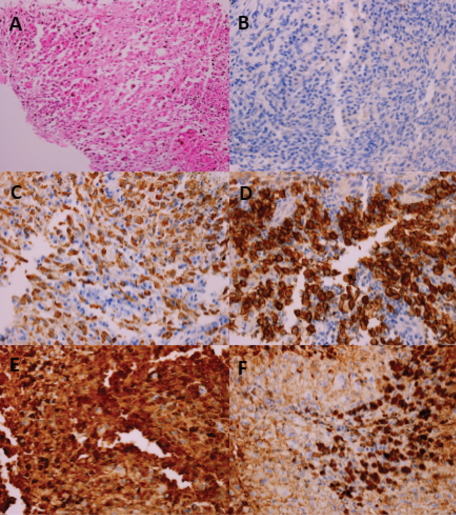
Figure 1: Intense fibrotic tissue with a dense infiltration with a large number of chronic inflammatory cell infiltration, mainly lymphocytes and plasma cells (A: Hematoxylin and eosin stain; original magnification ×200); Plasma cells were negative for CKAE1/AE3 body (B), and show intense and diffuse positive staining for CD38 (C), CD138 (D), IgG (E), while they are partially positive for IgG4 (F) (Immunochemistry stain, original magnification ×400).