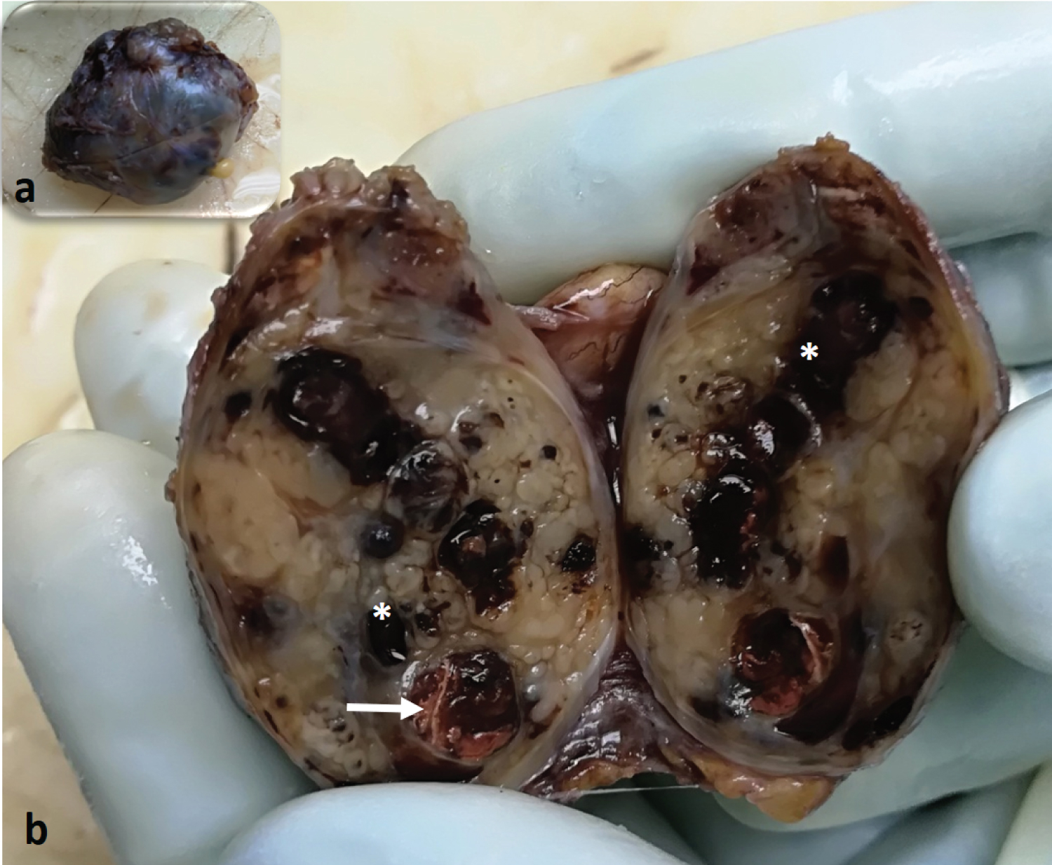
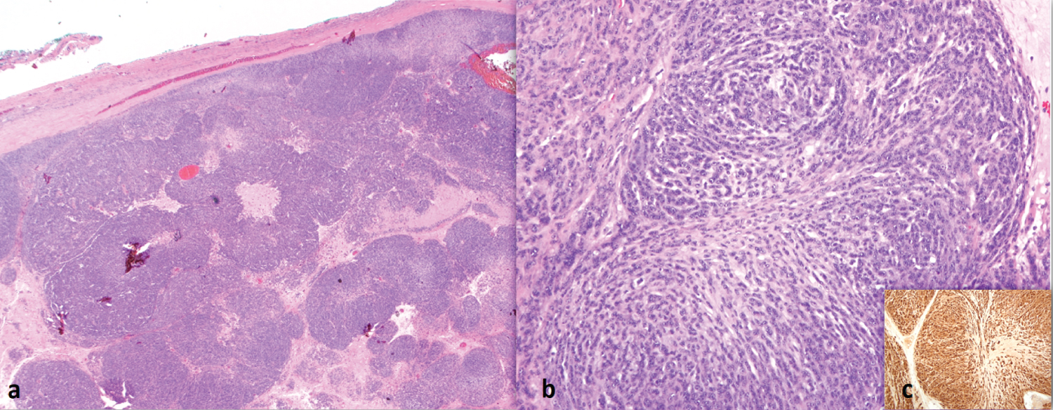
A 75-year-old man presented with a slow-growing patellar swelling with no history of pain or trauma to the site. The mass was clinically diagnosed as sebaceous cyst as it is well-circumscribed and located at the dermal-subcutanoeus tissue junction. Macroscopic cut section of the 40 mm, encapsulated mass showed variegated tan-yellowish cut surface with areas of haemorrhage and necrosis (Figure 1a and Figure 1b). Microscopically, it was well-circumscribed, composed of lobules of neoplastic cells separated by fibrous and hyalinised stroma. The neoplastic cells exhibit pleomorphic, round to oval, vesicular nuclei, prominent nucleoli and moderate amount of cytoplasm. Immunohistochemically, the neoplastic cells show strong and diffuse positivity for S100 (Figure 2a, Figure 2b and Figure 2c). They are negative for CK AE1/AE3, HMB45, Melan A, smooth muscle actin, desmin and CD34. Final diagnosis of epithelioid MPNST was made. The patient underwent radiotherapy following surgical excision and had no evidence of recurrence.
Epithelioid MPNST is rare variant of MPNST that is not associated with Neurofibromatosis type 1 (NF1) [1]. They may present at any site but cutaneous epithelioid MPNST commonly arise at the trunk and extremities. It has been reported to be slow-growing and may be painful. It has to be differentiated with other neoplastic entities showing epithelioid morphology for instance melanoma, epithelioid sarcoma, myoepithelial sarcoma and metastatic carcinoma [2,3]. Typical histologic features of epithelioid MPNST include multilobulated appearance with epithelioid cells arranged in cords and solid sheets. Epithelioid MPNST tends to shows strong and diffuse staining for S100, in contrast to conventional MPNST. When found at cutaneous location, it has the potential to pursue an unfavourable outcome compelling long-term follow-up [3].
The authors declare no competing interests.
Patient consent has been obtained.
There is no funding received for this submission.
All authors contributed equally to the work of this manuscript.

Figure 1: (a) Inset shows a lobulated mass with smooth outer surface; (b) Cut sections shows an encapsulated well circumscribed mass with lobulated, variegated tan-yellowish cut surface with areas of haemorrhage (asterisks) and necrosis (arrow).

Figure 2: (a) Microscopically, the mass is composed of lobules of neoplastic cells separated by fibrous and hyalinised stroma. The neoplastic cells are arranged in clusters and cords within the lobules with hypercellular periphery and less cellular, myxoid central region (H&E, 12.5x); (b) The neoplastic cells exhibit pleomorphic, round to oval, vesicular nuclei, prominent nucleoli and moderate amount of cytoplasm (H&E, 100x); (c) Inset shows the neoplastic cells are positive for S100.