Mitral Stenosis, Left Atrium, Spontaneous Echo Contrast
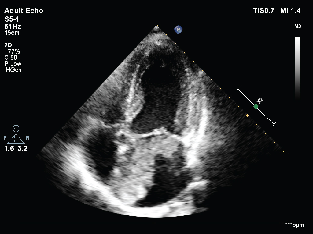
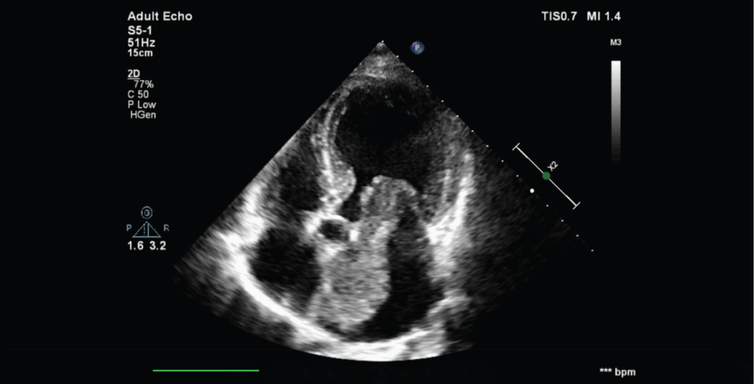
44-year-old male presented with shortness of breath for last 8 months. He had history suggestive of paroxysmal nocturnal dyspnoea. He had no history of rheumatic fever and had no complains of chest pain, palpitation or syncope. His ECG revealed no significant abnormalities, Auscultation revealed mid diastolic rumbling murmur best heard in the apical region. This murmur was changing in intensity and duration along with positional changes from standing to supine. Tumour plop was audible distinctly. Transthoracic Echocardiography (TTE) was done which revealed large echogenic structure attached to atrial septum protruding to left ventricle through mitral valve orifice. There was no echo contrast noted. No significant valvular abnormalities noted. Left atrial myxoma was diagnosed based on morphological findings and keeping in mind the absence of any embolic manifestations. Figure 1 showing large atrial myxoma attached to atrial septum. Figure 2 showing obstruction of mitral orifice by the tumour mass. Video 1 is the video of atrial myxoma moving through mitral valve orifice and protruding into left ventricle. This protrusion is causing obstruction in left ventricle filling which is the reason for symptoms similar to mitral stenosis. Myxoma is most common primary tumour of heart. The most common location is left atrium. Diagnosis of myxoma is challenging as patient may be asymptomatic. The symptoms mimic that of mitral stenosis as the tumour mass obstructs the filling of left ventricle leading to high atrioventricular gradient. Patients may presents with hemodynamic consequences of high left atrial pressure and/or venous congestion that includes dyspnoea, orthopnoea, PND, acute pulmonary oedema, lower limb oedema, hepatomegaly. Embolic complications represent a serious complication of myxoma. Migration of the tumour or its fragmentation, or even the posting of thrombi and vegetations adherent to the tumour surface may be responsible for such occurrences. Therefore early diagnosis is very crucial for proper management.
None.
None.

Figure 1: Large left atrial myxoma attached to interatrial septum.

Figure 2: Showing large left atrial myxoma obstructing mitral orifice.
Video 1: Showing large LA myxoma attached to atrial septum protruding into left ventricle obstructing the mitral orifice.