Mitral Stenosis, Left Atrium, Spontaneous Echo Contrast
24-year-old male with very severe mitral stenosis of rheumatic etiology presented with one episode of transient left sided hemiparesis with full recovery within 24 hours. He was on optimum medical management that includes diuretics, beta blocker and digoxin. Her heart rate was 62/minutes and BP was 110/64 mm of Hg. She was also receiving injection benzathine penicillin once every three weeks. Transthoracic Echocardiography (TTE) was done which revealed thickened anterior and posterior mitral leaflet with restricted mobility. Mitral valvular area was 0.636 cm2 in 2D planimetry. Left atrium was dilated with measurements of 54 ml/m2 in volume. She was planned for PTMC (Percutaneous transluminal mitral commissurotomy) and TEE (transesophageal echocardiography) was planned for further evaluation. TEE revealed LA Spontaneous Echo Contrast (SEC) or smoke in the left atrium. It mandates initiation of oral anticoagulation in this patient. Coagulation profile was advised after initiation of oral warfarin therapy and titrations of doses were done to achieve proper INR value. Figure 1 demonstrating the LA smoke in swirling pattern in TEE (Left panel) and mitral stenosis in TTE in right panel. Video 1 showing the same in video format which is clearly showing the LA smoke of grade 2+ to 3+ intensity. Left Atrial (LA) appendage peak emptying flow velocity (LAAEV) was found to be 15 cm/s. This explains the occurrence of embolic manifestations in the patient. Therefore, TEE examination has a special role in such situations even when TTE is not showing any such abnormalities.
None.
None.
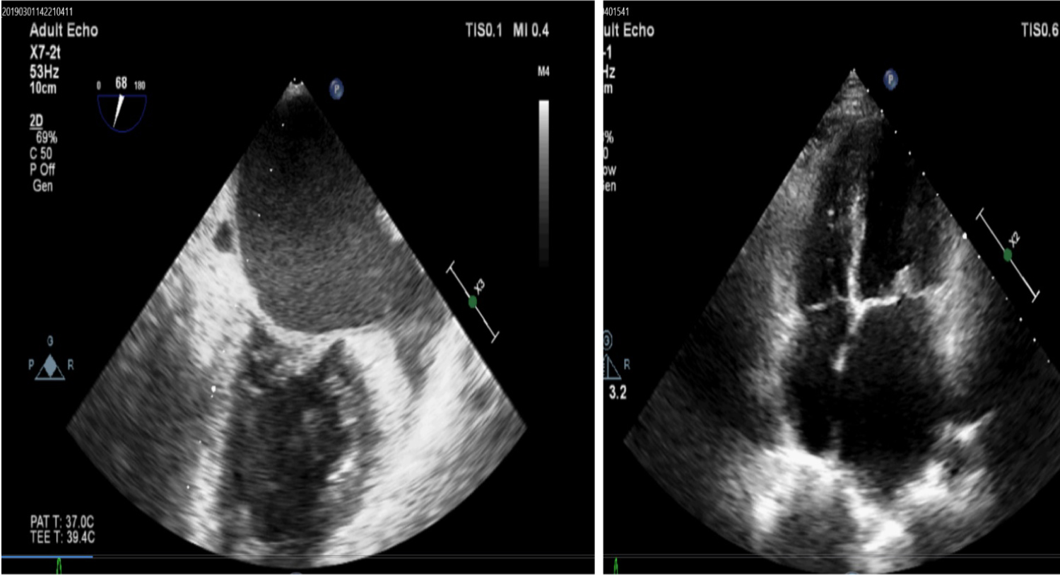
Figure 1: Showing the LA smoke in swirling pattern in TEE (Left panel) and mitral stenosis in TTE in right panel.