LA (Left Atrium) Clot, TTE (Trans Thoracic Echocardiography)
43-year-old lady presented with shortness of breath for past 7 months. She had history suggestive of Paroxysmal Nocturnal Dyspnoea (PND). She had also few episodes of palpitations. She had no history of rheumatic fever in the past. On physical examination mid diastolic rumbling murmur was audible at the apex which changed with posture. There was no history of fever and/or any embolic manifestations. Electrocardiogram revealed left atrial abnormalities. Transthoracic Echocardiogram was done which revealed severe mitral stenosis and mild mitral regurgitation. It was noted that a large ball of clot which is well organized moving inside the left atrium freely (Figure 1 and Video 1). This type of free ball clot is rare now a days. She was started on anticoagulation immediately and appropriate therapy.
None.
None.
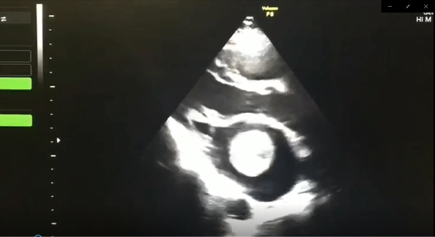
Figure 1: Transthoracic echocardiogram.