A ureterocele is a cystic dilatation of distal ureter in the bladder, urethra or both [1]. Commonly, ureterocele is found in the paediatric population due to regular antenatal scanning. The incidence of ureterocelesis reported as 1 in 500 [2], and 4 to 6 times as common in females as males [3]. Although ureteroceles in adults may be symptomatic, they are usually incidental findings.
We present an interesting magnetic resonance imaging (MRI) image of a left ureterocele of a 62-year-old man who had an MRI prostate to investigate his raised PSA. Classically, this is known as a cobra head sign.
Ureterocele, Cobra head sign
A 62-year-old man was referred by his general practitioner to the urology outpatient clinic as his routine blood test showed a raised prostate-specific antigen (PSA) of 10 ug/l. On further questioning, he has minimal lower urinary tract symptoms and there was no symptom to suggest that he had a urinary tract infection recently. Examinations of his abdomen and male genitalia including a digital rectal examination were normal. As recommended by the latest National Institute for Health and Care Excellence (NICE), he underwent a MRI prostate. Not only his MRI scan has picked up a Prostate Imaging Reporting and Data System (PI-RADS) 3 lesion in his anterior part of his prostate, but it has also picked up an incidental left ureterocele which classically is known as a cobra head sign (Figure 1).
In light of the lesion on his MRI prostate, he underwent a template biopsy of his prostate which fortunately was uneventful. Histologically, none of his 43 prostate biopsy cores picked up any malignancy. His case was subsequently discussed in the local multidisciplinary meeting and the decision from the team was to monitor his PSA in the community.
With regards to left ureterocele, as he was completely asymptomatic, he did not undergo further investigation or treatment for it.
N/A.
None.
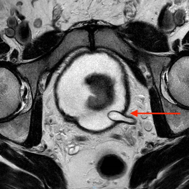
Figure 1: Cobra head sign of the left ureterocele.