Cavernous hemangioma, Spine, Cord compression, Vascular malformation
Cavernous hemangioma is a common vascular lesion. These tumors usually appear on the skin [1]. Vertebral hemangioma is asymptomatic, common, and benign lesion with an incidence of 10-12% and can cause acute spinal cord compression. Vertebral hemangiomas can become symptomatic in rare cases (about 1%). This lesion mostly occurs in the body of vertebral bones. There have been few case reports of spinal cord hemangioma so we intend to report some interesting and rare images on this topic [1-3].
The patient is a 40-year-old married man was referred with the complaint of low back pain. The pain was referred to proximal of lower limbs. Past medical history was negative. The cranial nerves exam were normal. The first diagnosis was a discal hernia. After lumbar MRI, a thickening was determined in the upper section. Therefore, another MRI with gastrographine was done for upper parts. A mass was detected in spinal foramen. Following surgery, the extradural lesion without extension to vertebrae was detected. The pathology report was the cavernous hemangioma.
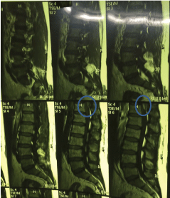
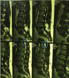
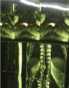
Extradural cavernous hemangiomas are usually isointense on T1W (Figure 1) and hyperintense on T2W images (Figure 2) and show homogenous contrast enhancement (Figure 3) because of the presence of sinusoidal channels.
Cavernous hemangiomas are benign vascular abnormalities. Aggression of hemangiomas into the epidural and foraminal space is rare and can cause soft tissue compression. Actually, Neurologic deficit and is one of the rare complication of this lesion [1,3]. In our case, a mass was detected in spinal foramen and it was the cause of the low back pain.
Metastatic disease and primary bony malignancy are the most important differential diagnosis of this disease. Surgery is a good treatment strategy. But there is huge concern about following bleeding [2-4]. In our case, surgery was done and it was successful.
They are so many benign vascular lesion and often involve soft tissue mostly during childhood. Differential diagnosis of this condition is so wide but tumors are the most important.
We want to thank patient's cooperation.
No.
Seyed Ahmad Mirhosseini and Mohammad Ebrahim Ghanei prepared images and gathered data and Reza Bidaki and Ehsan Zarepur wrote the primary draft, revised and submitted it.

Figure 1: Sagittal T1-weighted image shows an iso signal extra-medullary extra-dural soft tissue mass in right side of the dorsal spinal canal at T11 & T12 vertebral levels, causing thecal sac compression.

Figure 2: Sagittal T2-weighted image showing high signal intensity in the mass lesion.

Figure 3: Axial (superior) sagittal (left) and coronal post-contrast, T1-weighted images show intense homogenous enhancement of the epidural lesion with moderate extension into the right foramina (red arrow).