We report a large fistula between left main coronary artery and pulmonary trunk as major source of pulmonary blood flow in a patient with pulmonary atresia and ventricular septal defect.
Aortopulmonary collateral, Coronary to pulmonary collateral, Coronary fistula
Pulmonary atresia with ventricular septal defect is associated with variable aortic to pulmonary arterial connections. Coronary artery to pulmonary artery collaterals are rare sources of pulmonary blood flow, and when present, they are associated with other major aortopulmonary collaterals, and will have no significant proximal coronary artery dilatation [1,2].
We report a 21-year-old female patient with a diagnosis of pulmonary atresia and ventricular septal defect. She had no interventions done previously. She presented with severe cyanosis, exercise intolerance, clubbing, and polycythemia. Echocardiography showed a large ventricular septal defect, overriding aorta, pulmonary atresia and good sized central pulmonary artery. CT angiography showed a well developed pulmonary trunk, normal size left pulmonary artery and a small right pulmonary artery. There was severely dilated left coronary sinus and left main coronary artery (Figure 1). Cardiac catheterization was done to study the anatomy of pulmonary blood flow, and to measure pulmonary arterial pressure.
Aortic root angiography showed severely dilated left coronary sinus and left main coronary artery, with a tortuous, spiral-like fistulato pulmonary trunk that comprised most of pulmonary blood flow. Additional small aortpulmonary collateral was seen from descending aorta to right lung. Distal left coronary artery appeared normal in size, although underfilled due to steal. Right coronary artery was normal (Figure 2). Pressure measurement was done by a microcatheter manipulated through the fistula to the pulmonary trunk, showing a pressure of 88/55 Hg. Calculated pulmonary vascular resistance was 25 Woods Units with no significant response to oxygen, precluding surgical repair.
Supplementary Video S1: Aortic root angiogram in a patient with pulmonary atresia and ventricular septal defect demonstrating the large, tortuous fistula between the dilated left main coronary artery and the pulmonary trunk. Distal to the origin of the fistula, the left coronary artery is underfilled due to steal. Pulmonary trunk is well developed with confluent branch pulmonary arteries.
This research received no grant from any funding agency, commercial or non-for-profit.
None.
This report does not involve human or animal experimentation.
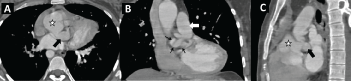
Figure 1: CT angiogram for a patient with pulmonary atresia and ventricular septal defect demonstrating a large, tortuous fistula (small arrows) between the left coronary artery and the pulmonary trunk as the main source of pulmonary blood flow. The aortic root (star) is dilated, the left main coronary artery is severely dilated (black arrow).

Figure 2: Aortic root angiography for a patient with pulmonary atresia and ventricular septal defect (A) A hydrophilic wire was passed from the aortic root across the left coronary artery to the pulmonary trunk, wire course demonstrating the spiral course of the fistula; (B) Aortic root angiogram showing a severely dilated left main coronary artery (white arrow); (C) Schematic showing the cardiac structures of interest.
AO: Ascending Aorta; LCA: Left Coronary Artery; PA: Pulmonary Artery; RCA: Right Coronary Artery.
Supplementary Video S1: Aortic root angiogram in a patient with pulmonary atresia and ventricular septal defect demonstrating the large, tortuous fistula between the dilated left main coronary artery and the pulmonary trunk. Distal to the origin of the fistula, the left coronary artery is underfilled due to steal. Pulmonary trunk is well developed with confluent branch pulmonary arteries.