Coronary artery ectasia (CAE) is defined as dilatation of the coronary artery one and a half times greater than that of an adjacent normal segment [1]. It usually accompanies coronary artery disease (CAD). Our objective in this study was to examine the clinical characteristics of CAE and its prognosis.
We recognized CAE in five patients between February 2016 and June 2016 admitted to Rashid Hospital, Dubai, United Arab Emirates. The patients presented with chest pain and were diagnosed as acute coronary syndrome (ACS).
A 29-year-old male presented with central chest pain consistent with cardiac ischaemia. His past medical history was unremarkable. He had never smoked and no other risk factors for CAD were identified. Physical examination was unremarkable. Inferior ST-segment-elevation was demonstrated on an electrocardiogram (ECG) and fibrinolysis was administered. Due to inadequate ST-segment-elevation resolution he proceeded for urgent coronary angiography with potential for rescue percutaneous coronary intervention (PCI). This revealed CAE of the right coronary artery (RCA) with thrombus (Figure 1A). Reduced coronary flow was present with Thrombolysis in myocardial infarction (TIMI) 2 flow. In addition there was CAE of the left anterior descending (LAD) coronary artery also with TIMI 2 flow (Figure 1B). Thrombus aspiration was performed and TIMI 3 flow was established (Figure 1C). Intravenous glycoprotien IIb/IIIa was administered. No stent was deployed however as no flow-limiting lesion was present and due to the potential for malposition if a stent was to be deployed in an ectatic segment. The peak serum troponin T was 1.93 ng/mL (ULN 0.01 ng/mL) the day following presentation. Echocardiography demonstrated mild impairment of left ventricular systolic function with mid inferior and mid inferoseptal hypokinesis. Standard medical therapy was continued for ACS. The remaining hospital course was uneventful and he was discharged on the fourth day following admission. Dual antiplatelet therapy was suggested for twelve months and he was discharged on guideline recommended therapy.
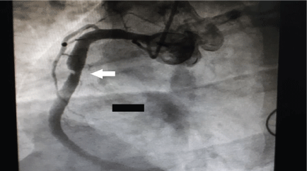 Figure 1A: Initial angiographic findings. Right coronary artery ectasia with thrombus at mid right coronary artery (white arrow). View Figure 1A
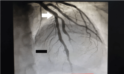 Figure 1B: Angiographic findings. Ectasia of left anterior descending coronary artery (white arrows). View Figure 1B
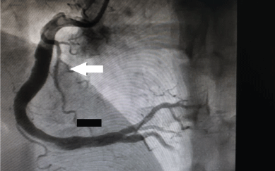 Figure 1C: Follow up angiographic findings after aspiration of mid right coronary artery thrombus (white arrow). View Figure 1C
A 44-year-old male with no known risk factors for CAD presented with typical central chest pain. Physical examination was unremarkable. ST and T wave changes were demonstrated in the inferior leads on an ECG. The peak serum troponin T was 39.19 ng/mL (ULN 0.01 ng/mL) the day following presentation. Guideline recommended ACS management was commenced. Coronary angiography was undertaken and revealed CAE in all epicardial vessels but more evident in the RCA (Figure 2A) with TIMI 2 flow in all vessels (Figure 2B). No flow limiting lesions were identified however and accordingly medical management was continued. Mild impairment of left ventricular systolic function was demonstrated on echocardiography with mid inferior hypokinesis. The rest of his hospital stay was unremarkable and he was discharged on day four on guideline recommended medical therapy in addition to dual antiplatelet therapy for twelve months.
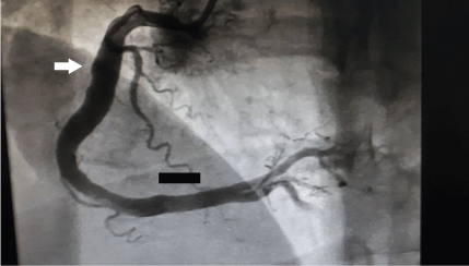 Figure 2A: Angiographic findings. Ectasia of the right coronary artery (white arrow). View Figure 2A
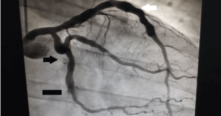 Figure 2B: Angiographic findings. Ectasia of left anterior descending (white arrow) and left circumflex coronary arteries (black arrow). View Figure 2B
A 41-year-old male, with an unremarkable past medical history apart from smoking, presented with typical central chest pain. Physical examination did not reveal any abnormal findings. Inferior ST-segment-elevation was demonstrated on an ECG and the patient proceeded to coronary angiography with the potential for primary PCI. The coronary angiography demonstrated right CAE with a distal thrombotic occlusion (Figure 3A). The proximal segment of the LAD was also ectatic with TIMI 2 flow (Figure 3B). Thrombus aspiration and stent deployment was performed (Figure 3C). The peak serum troponin T was 13.43 ng/mL (ULN 0.01 ng/mL) the day following presentation. Echocardiography demonstrated akinesia of the mid basal inferior and mid lateral walls with moderate impairment of left ventricular systolic function. Standard medical therapy was continued and the rest of his hospital stay was uneventful.
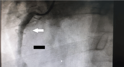 Figure 3A: Angiographic findings. Ectasia of right coronary artery with distal thrombotic occlusion (white arrow). View Figure 3A
 Figure 3B: Angiographic findings. Ectasia of proximal left anterior descending coronary artery (white arrow). View Figure 3B
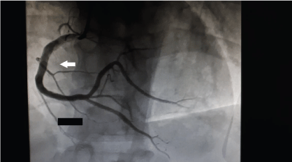 Figure 3C: Angiographic findings. Thrombus aspiration and stent deployment was performed to the right coronary artery (white arrow). View Figure 3C
A 49-year-old male, with no significant past medical history, presented with typical central chest pain. Physical examination was unremarkable. ST-segment-changes were demonstrated on the inferior leads of his ECG. The peak serum troponin T was 1.6 ng/mL (ULN 0.01 ng/mL). Standard medical therapy was continued for the ACS and coronary angiography was undertaken. This revealed CAE in the RCA but with TIMI 3 flow (Figure 4A and Figure 4B). The other epicardial coronary arteries were not affected by CAE. A subsequent echocardiogram demonstrated preserved left ventricular systolic function. The patient was continued on medical therapy that included dual antiplatelet therapy and he was discharged following a three day stay in hospital.
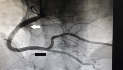 Figure 4A: Angiographic findings. Ectasia of right coronary artery (white arrow). View Figure 4A
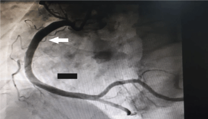 Figure 4B: Angiographic findings. Ectasia of right coronary artery (white arrow). View Figure 4B
A 29-year-old male smoker, with no significant past medical history, presented with typical central chest pain. Physical examination was unremarkable. Inferolateral ST-segment changes were demonstrated on an ECG. The peak serum troponin T was 0.16 ng/mL (ULN 0.01 ng/mL) the day following presentation. Standard medical therapy was commenced for ACS and coronary angiography was undertaken. This revealed CAE of a large obtuse marginal branch of the left circumflex coronary artery. The obtuse marginal was also occluded due to thrombus (Figure 5A). The other major epicardial coronary arteries were not affected by CAE. Aspiration was performed that restored TIMI 3 flow and no severe stenosis was demonstrated (Figure 5B and Figure 5C). Hence a stent was not deployed. A subsequent echocardiogram demonstrated preserved left ventricular systolic function but with akinesis of the mid posterior wall. The rest of his hospital stay was uneventful and he was discharged on day three on medical therapy that included dual antiplatelets.
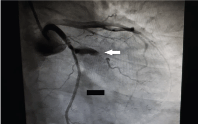 Figure 5A: Angiographic findings. Ectasia of obtuse marginal coronary artery with thrombus (white arrow). View Figure 5A
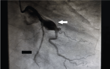 Figure 5B: Angiographic findings. Ectasia of obtuse marginal coronary artery following thrombus aspiration (white arrow). View Figure 5B
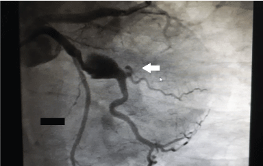 Figure 5C: Angiographic findings. Ectasia of obtuse marginal coronary artery after thrombus aspiration (white arrow). View Figure 5C
The prevalence of CAE in the literature varies between 1.2-6% [1]. Desmopoulos, et al. describe an association of traditional risk factors for CAD for CAE except diabetes [2]. No specific predisposing risk factor was identified however [2]. The prevalence of systemic hypertension was higher in some studies [1]. Sudhir, et al. found an increased prevalence of ectasia in familial hypercholesterolaemia but ectasia was not found to be related to age, hypertension, smoking or ethnicity [3]. Age seems to have no additional influence according to most investigators [2,3]. The limited case series we describe suggest lack of risk factors in those presenting with CAE. Two out of the five patients we presented were smokers however. In some previous studies the RCA was reported to be the most commonly involved vessel, whereas others reported the LAD to be more frequently involved [1,2,4,5]. The RCA was the most commonly involved vessel in our study.
The authors report different deals in coronary ectasia that could be construed as a conflict of interest.