Sarcoidosis is a multisystem inflammatory disease of unknown etiology, characterized by noncaseating granuloma variably infiltrating the respiratory tract, ganglions, eyes, internal organs and the skin. The heterogeneity of cutaneous sarcoidosis represents a diagnostic challenge for physicians and affirms its reputation as a "great imitator". Common specific lesions associated with sarcoidosis are papules, plaques, nodules, scar sarcoidosis and lupus pernio. Atypical presentations of sarcoidosis do exist and include ichthyosiform, alopecic, erythrodermic, angiolupoid and verrucous variants. Verrucous sarcoidosis is an extremely rare manifestation. Previous reports of this phenotype are limited to 16 cases, with only 5 previous reports of facial verrucous sarcoidosis reported in the literature. Here we report a case of a 50-year-old Moroccan woman who had an unusual cutaneous presentation of facial verrucous sarcoidosis.
Sarcoidosis is a systemic disease characterized by the formation of noncaseating granulomas in several organs. Various cutaneous manifestations of sarcoidosis occur in approximately 25% of patients and may indicate onset of the disease [1]. The heterogeneity of cutaneous sarcoidosis represents a diagnostic challenge for physicians and affirms its reputation as a "great imitator". Common specific lesions associated with sarcoidosis are papules, plaques, nodules, scar sarcoidosis and lupus pernio. Atypical presentations of sarcoidosis do exist and include ichthyosiform, alopecic, erythrodermic, angiolupoid and verrucous variants [2]. Verrucous sarcoidosis is an extremely rare manifestation and previous reports of this phenotype are very limited.
A 50-year-old Moroccan caucasian female with no history of systemic sarcoidosis, presented with a 6-month history of a nodular verrucous lesion of 3 cm of diameter on the nose (Figure 1). The lesion was treated initially as leishmaniasis by injections of glucantium without improvement. The patient consulted a surgeon who performed a total lesion excision without prior skin biopsy, thinking it may be a squamous cell carcinoma. Histology revealed noncaseating granuloma giganto-cellular with a sparse lymphocytic infiltrate of the dermis (Figure 2). Asteroid or Schaumann bodies were not identified. Non-polarizable foreign material and nor infectious source were identified. The evolution was marked by a relapse (Figure 3) after 6 months: An erythematous patch appeared in the same localization of the first lesion. The diagnosis of sarcoidosis was established by the histological findings and after eliminating other pathological causes of skin lesion. An angiotensin-converting enzyme testing was performed which was positive (81 IU) and a systematization assessment was carried out. The patient had no systemic involvement and was treated with topical and systemic corticosteroids (0.7 mg/kg/day) with good clinical development after 03 months (Figure 4).
Variability in the cutaneous features of sarcoidosis affirms the condition's designation as one of dermatology's "great imitators" [2]. The diagnosis often requires a clinical presentation congruent with histologic features in addition to exclusion of confounding aetiologies [3]. Verrucous sarcoidosis (VS) is an extremely rare subtype [4], with only 16 cases previously reported [5]. It was first described by Irgang in 1950 [6]. The majority of reported cases involve black patients [4] with concomitant systemic disease, principally of the respiratory tract [3,7]. Two cases have been noted in white women, one without systemic involvement at the time of diagnosis [5], our patient also presented with disease limited to the skin. VS most commonly affects the lower extremities, although 5 cases have been reported on the face [5,8-10]. Most of previous cases describe improvement with topical, intralesional and systemic corticosteroids [3].
Our case confirms the variation of lesions in cutaneous sarcoidosis. VS could represent a diagnostic pitfall; it may be confused with other inflammatory and neoplastic skin disorders. We believe that this example of VS highlights the importance of always performing a skin biopsy before any surgical procedure. Careful clinical and pathological correlation led to the proper diagnosis and therapy in our case.
The authors declared no potential conflicts of interest.
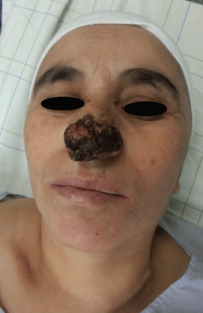
Figure 1: Nodular verrucous lesion of 3 cm of diameter on the nose.
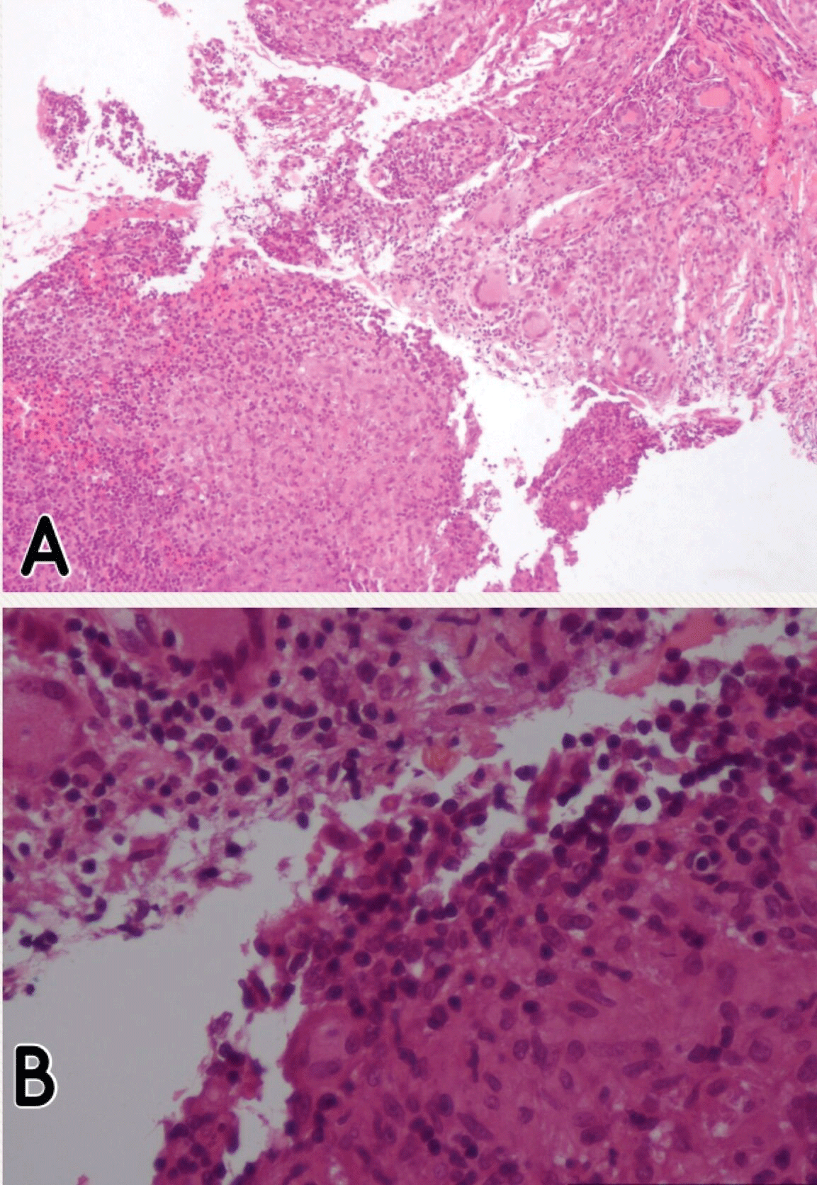
Figure 2: Noncaseating granuloma giganto-cellular with a sparse lymphocytic infiltrate of the dermis (A and B, Hematoxylin-eosinstain; original magnifications: A x40; B x100).
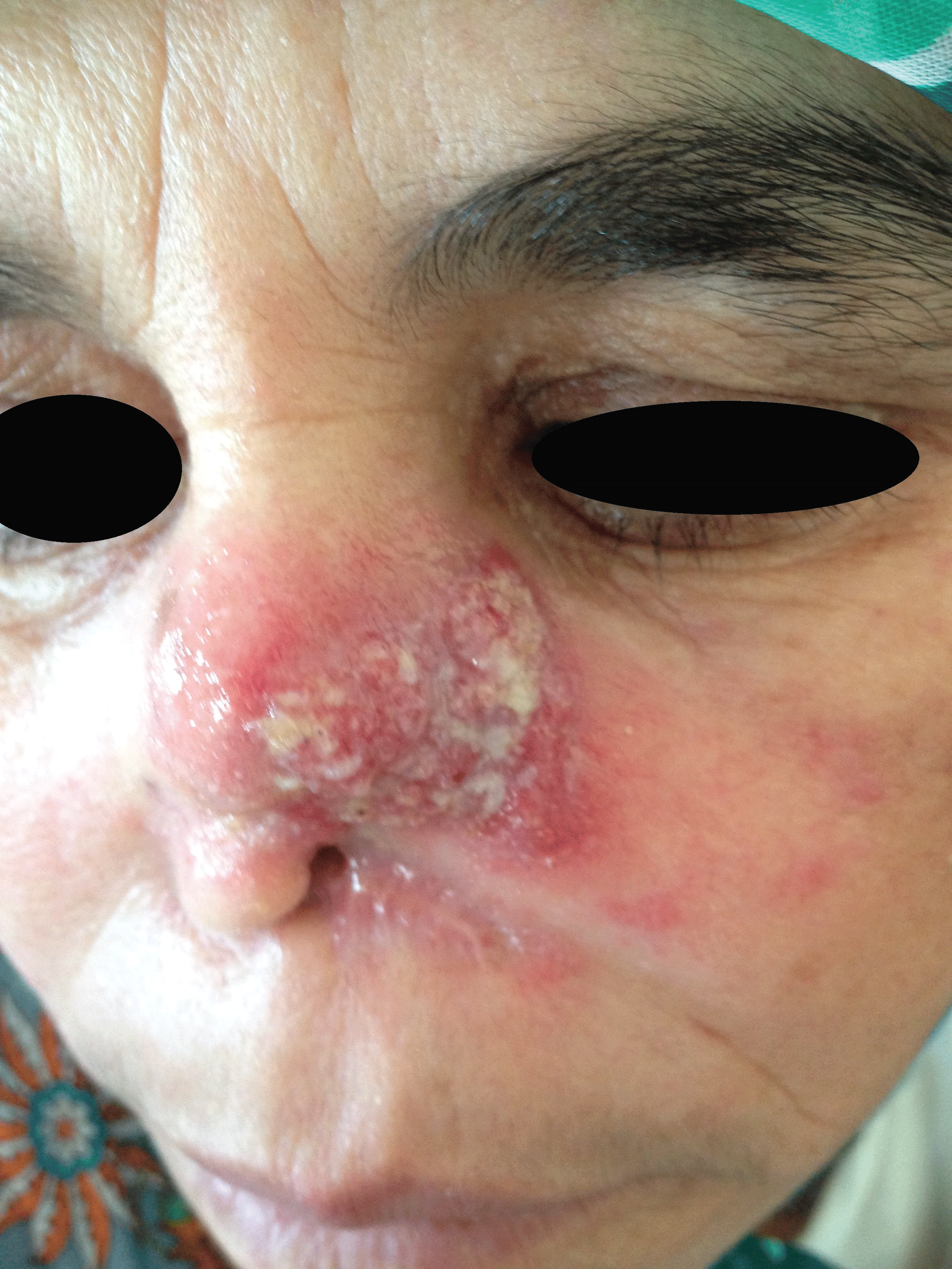
Figure 3: A relapse after 6 months: An erythematous patch appeared in the same localization of the first lesion.
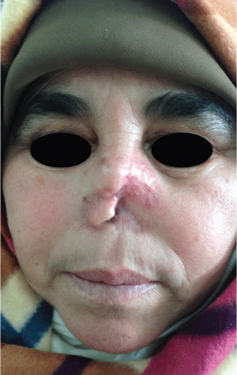
Figure 4: Clinical remission after 3 months of corticosteroid therapy.