Tarsal navicular bone dislocation is a rare condition which can lead to long term disability, mainly due to difficulties to obtain an anatomical reduction. Only few cases have been reported.
We report a case of a 21-year-old Cameroonian male who was admitted to the emergency department after a road traffic accident. The physical examination reveals a patient unable to bear weight, with a painful swelling of the antero-medial dorsal region of the right foot, without neurovascular deficits or another lesion. The plain X-ray and the CT scan of the right foot and ankle reveal a medial dislocation of the talonavicular joint without associated fracture. An open reduction was done and two Kirschner wires of 2 mm were place to maintain the reduction. The patient was discharge after five days with an ankle/foot X-ray showing successful reduction of the talonavicular joint. He was reviewed at one and twelve weeks later with no complain and a normal X-ray.
Tarsal navicular bone dislocation is a rare condition which can lead to long term disability. Even in resources constraint settings, a CT-scan should be done when available, in addition to radiography to rule out an associated lesion which can be missed by the X-ray, and to plan the surgical approach.
Navicular bone, Dislocation, Internal fixation, Open reduction
Isolated medial dislocation of the tarsal navicular bone without fracture is known to be a rare condition which can lead to long term disability, mainly due to difficulties to obtain an anatomical reduction and, degenerative bone lesions [1,2]. Only few cases have been reported in the literature [3,4]. Herein, we report the case of 21-year-old man with isolated right tarsal navicular bone dislocation. We highlight the diagnostic role of computed tomography (CT) scan to diagnosis and plan management of Chopart joint dislocations.
A 21-year-old Cameroonian male patient with no significant past history, presented at the emergency department with swollen and painful right foot following a road traffic accident one hour prior to consultation. The patient was the only passenger of a motorbike on a public road. He fell to his right side after the bike was pushed to the ground on its left by a car. The right foot caught in the rear wheel of the bike sustained a forced internal rotation, supination and adduction.
On examination, the patient was conscious with normal vital signs. The dorsal anteromedial region of the right foot was tender and swollen, without signs of neurovascular deficits or compartment syndrome. The rest of the physical examination was unremarkable.
A right foot/ankle X-ray requested and a CT scan showed a medial dislocation of the talonavicular joint without fracture (Figure 1a and Figure 1b).
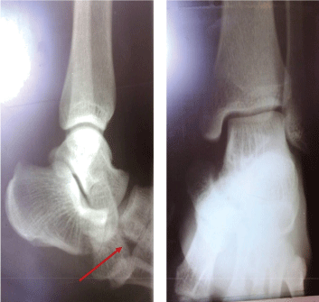 Figure 1A: An X-ray view of the right Ankle showing a medial luxation of the talonavicular joint (red arrow).
View Figure 1A
Figure 1A: An X-ray view of the right Ankle showing a medial luxation of the talonavicular joint (red arrow).
View Figure 1A
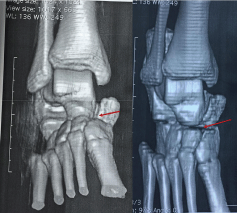 Figure 1B: A CT scan view of the right ankle/foot showing a medial luxation of the talonavicular joint.
View Figure 1B
Figure 1B: A CT scan view of the right ankle/foot showing a medial luxation of the talonavicular joint.
View Figure 1B
The decision to do an open reduction and fixation of the navicular bone was taken. Peroperatively, under spinal anesthesia, a 4 cm incision was made over the mid anterior face of the ankle, exposing the talonavicular joint (Figure 2). After flexing the knee, a medial pressure was applied on the scaphoid and a traction done longitudinally on the bone with a holding forceps, obtaining the reduction which was maintained in place with two 2 mm Kirschner wires. The subcutaneous tissue and skin were closed.
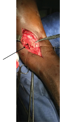 Figure 2: An operative view of the right ankle showing the absence of the navicular bone in the place normally field by it.
View Figure 2
Figure 2: An operative view of the right ankle showing the absence of the navicular bone in the place normally field by it.
View Figure 2
Postoperative day five was uneventful and a control radiograph showed anatomical reduction of the navicular bone (Figure 3). The patient was discharge on analgesics, and was advised not to bear weight on the affected foot. A follow-up visit was scheduled one week later, during which the patient was pain free and the physical examination was unremarkable. Another follow up visit was scheduled twelve week later the patient had no complaint, the physical examination was normal and the X-ray was unremarkable (Figure 4). We therefore plan to removed Kirschner wires after seven weeks, but unfortunately, the patient was see after 12 weeks and after that he was lost to follow up.
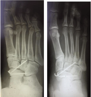 Figure 3: Post-operative X-ray at day 5 showing a normal talonavicular joint, with two K-wires maintaining the navicular bone in place.
View Figure 3
Figure 3: Post-operative X-ray at day 5 showing a normal talonavicular joint, with two K-wires maintaining the navicular bone in place.
View Figure 3
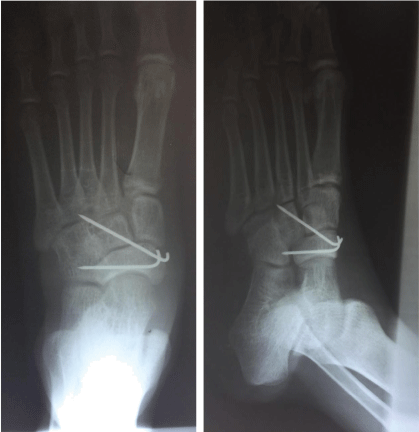 Figure 4: Post-operative X-ray done after 12 weeks showing a normal talonavicular joint, with two K-wires maintaining the navicular bone in place.
View Figure 4
Figure 4: Post-operative X-ray done after 12 weeks showing a normal talonavicular joint, with two K-wires maintaining the navicular bone in place.
View Figure 4
Isolated traumatic close medial dislocation of tarsal navicular bone is a rare lesion [2,4-10]. Usually, dislocation of the navicular bone is associated with the fracture of this bone or with the fracture of another tarsal bones, so much so that it has been suggest by Vaishya, et al. that, dislocation of the navicular bone could not occur without associated fracture [11], because of the rigidity of the ligaments and bones surrounding the navicular bone [10,11]. Several mechanisms have been described as hyper-plantar flexion and axial loading or abduction pronation [11,12]. In our case, the mechanism described is not the one usually recognized, since it was a forced plantar flexion and adduction. But this stand on patient report and, only biomechanical study or injury recording can provide unquestionable evidence.
Since the appropriate treatment of this lesion is not clearly established [1,9,11], and that we did not have a per operative image intensifier to evaluate the quality of the reduction we initially opted for an open reduction under anaesthesia, which was successfully done and, the navicular bone maintains in place with two K-wires with satisfactory results. Such lesions are not frequent and there is a lack of therapeutic guidelines, therefore there is a need to report any single case to pool enough experience that can establish consensus on the treatment of navicular bone dislocations.
In this case the choice of K-wires instead of screws to maintain the reduction was done with satisfactory results. But other surgeon may have chosen stronger fixation mean as screws to allow early weight bearing.
A CT-scan was done to rule out an associated fracture in our case. Even in resource constraint settings, a CT-scan should be done when available in addition to radiography to rule out any associated lesion which can be missed on X-ray, and may help to plan a more complete treatment of various lesions [9].
The follow up of patients with dislocation of tarsal navicular bone should be done keeping in mind the possibility of necrosis or chondrolysis that occur in the case reported by Dias, et al. and that prompted an arthrodesis after 6 weeks with satisfactory result 13 years later [12].
Tarsal navicular bone dislocation is a rare condition which can lead to long-term disability. Where available, a CT-scan should be done in addition to an X-ray to rule out an associated lesion which can be missed by the X-ray, and to plan the surgical approach.
A written informed consent was obtained from our patient for publication of this case report and accompanying images.
CD drafted the manuscript. CD and GML revised the manuscript for methodological and intellectual content. NYMA, TBJD, DF and IF critically revised the manuscript for intellectual content. GML is the guarantor of the Case. All authors approved the final version of this manuscript.
None.
None.
No additional data are available.