Primary paraurethral tumours of benign nature in females are quite infrequent. Physical examination and imaging modalities like ultrasound and magnetic resonance imaging of the pelvis help in characterising the mass morphologically and structurally. They also help to decipher the relationship of the mass with the urethra and the vagina.
A 51-year-old parous lady was referred to our urogynaecology clinic in view of a long standing vaginal lump. On examination, she had a 3 × 3 cm firm, nontender, mobile anterior vaginal mass with a smooth surface. The upper limit of the lump was about one cm below the urethral orifice. An ultrasound of the pelvis revealed a 3.1 × 2.5 × 1.5 cm well circumscribed hypervascular hypoechoic mass with a vascular pedicle at the region of the introitus. She was counselled for excision of the vaginal mass. Intraoperatively, a metal urethral catheter was inserted. A midline vertical incision was made over the mass and it was enucleated. A check cystourethroscopy performed was normal. The tumour bed and the vaginal mucosa were closed using vicryl sutures.
The patient recovered well. The histopathology report revealed a smooth muscle tumour with epithelioid and spindle cells arranged in cords, nests and fascicles.
Epithelioid leiomyomas are very rare. Surgical excision and histopathologic examination is generally required to confirm the diagnosis and to exclude the presence of a sarcoma. Long term follow up is advised as late recurrences may occur.
Primary paraurethral tumours of benign nature in females are quite infrequent. A tumour in this area may present with symptoms like a lump coming out of the vagina, dysuria, dyspareunia or retention of urine, which might erroneously be thought to be due to an uterovaginal prolapse. A physical examination of the lump helps in arriving at a diagnosis of vaginal tumour [1-3]. Different imaging modalities like ultrasound and magnetic resonance imaging of the pelvis help in characterising the mass morphologically and structurally. They also help to decipher the relationship of the mass with the urethra and the vagina. These imaging studies are helpful to plan a surgical excision of the mass which can be performed either through a transvaginal or transvesical approach depending upon its location [2]. Herein, we report a case, where the patient presented to our clinic with the complaint of a vaginal lump and was later diagnosed to have a anterior vaginal wall leiomyoma.
A 51-year-old parous lady was referred to our urogynaecology clinic in view of a long standing vaginal lump. She complained of a painless lump coming out of the vagina for 7 to 8 years, gradually increasing in size and occasional urinary urgency which was not bothersome. On examination, she had a 3 × 3 cm firm, nontender, mobile anterior vaginal mass with a smooth surface. The upper limit of the lump was about one cm below the urethral orifice.
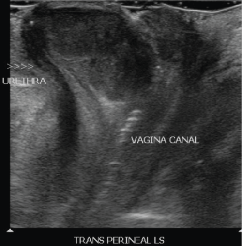
Urine cytology was negative for malignant cells and an ultrasound study of the kidneys and urinary bladder was normal. An ultrasound of the pelvis revealed a 3.1 × 2.5 × 1.5 cm well circumscribed hypervascular hypoechoic mass with a vascular pedicle at the region of the introitus. The vascular stalk appeared to be extending from the vagina. It was seen separate from the urethra. Transperineal ultrasound study was also performed. Overall features were nonspecific and indeterminate. It was suggested that it may be arising from the vagina or could be a prolapsed polyp. The patient was offered two management options - either conservative with a repeat ultrasound pelvis in a few months' time or surgical excision of the vaginal lump. She opted for conservative management.
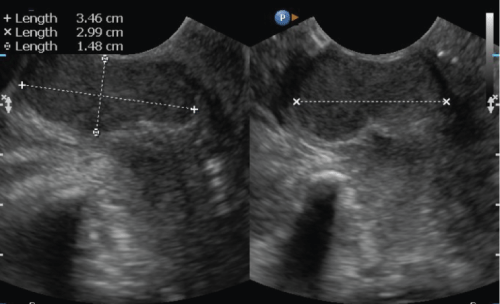
A repeat ultrasound pelvis three months later revealed a 1 × 0.9 cm endometrial polyp which was not present in the previous study. The anterior vaginal wall mass was largely unchanged from the previous study and now measured 3.5 × 3 × 1.5 cm (Figure 1 and Figure 2). It was again seen separate from the urethra. She was counselled for excision of the vaginal mass and a diagnostic hysteroscopy, polypectomy and endometrial curettage. She consented for the procedure. A metal catheter was inserted into the urethra during the procedure to avoid damage to it during enucleation of the mass. Intraoperatively, a midline vertical incision was made in the anterior vaginal wall over the mass, starting one cm below the urethral orifice and the mass was enucleated. The margins of the mass were very close to the urethral outlet. A check cystourethroscopy performed was normal. The tumour bed was closed using 2-0 vicryl suture and the vaginal mucosa was closed using 2-0 vicryl rapide suture.
 Figure 1: Ultrasound picture of anterior vaginal wall mass. View Figure 1
 Figure 2: Transperineal view of the mass and it's relation to the urethra. View Figure 2
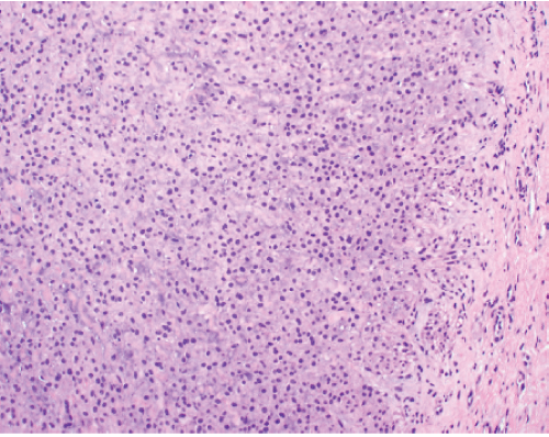
The patient recovered well. The indwelling catheter was removed the next morning and she was able to pass urine well with minimal residual urine on bladder scan. She was discharged on the first postoperative day. The histopathology report revealed a smooth muscle tumour with epithelioid and spindle cells arranged in cords, nests and fascicles (Figure 3). There were no mitotic figures or atypia. Immunohistochemistry showed reactivity for SMA (Smooth Muscle Actin), desmin and caldesmon and presence of estrogen and progesterone receptors. In view of rarity of vaginal leiomyomas, the patient is being followed up.
 Figure 3: A nodular lesion with well-defined border showing an area of epithelioid cells arranged in cords and nests (H & E, X200). View Figure 3
A vaginal tumour may be mistaken for several other benign conditions of the vagina, including uterovaginal prolapse, vaginal cysts, urethral diverticulum and urethral or vaginal malignancy [4-7]. Vaginal fibroids are rare and only 300 cases have been reported in literature. Of all the solid tumours occurring in the vagina, 4.5% were reported to be leiomyomas in a survey [8].
The first report of a vaginal leiomyoma was made by Denys de Leyden in 1733 [9]. It generally occurs in 35-50 year-old women and characteristically arises from the anterior vaginal wall in the midline [10]. It is more commonly found in Caucasian women. Although the origin is not clear, it most likely arises from local artery musculature and embryonal rests [9]. It typically appears to be a firm, non-tender, non-fluctuant vaginal mass with a smooth surface and no associated discharge on physical examination. It may be asymptomatic to begin with due to the elasticity of the vagina but may cause bladder outlet obstruction or mass related symptoms [8].
The American College of Radiology recommends ultrasound as the first-line imaging modality for vaginal masses [11]. Its main drawbacks are operator dependence and the fact that the transducer may bypass the perineum or vagina when inserted into the anterior or posterior fornix. If the imaging is inconclusive or non-diagnostic, a pelvic MRI is recommended [12].
Surgical excision via a vaginal approach is the preferred treatment, since the specimen can be thoroughly examined, and foci of malignant degeneration can be excluded [9,10]. The use of a urethral catheter during the procedure may help in tissue dissection and aid in preventing and identifying urethral injury. If skeletonization of the urethral and bladder support occurs due to dissection, a colporrhaphy/pubourethral plication must be performed [9]. The abdominal approach for excision should only be resorted to if the lesion is unapproachable via the vaginal route or if it involves the vaginal fornices [4].
Epitheloid leiomyomas of the genital tract are very rare [13]. Tavassoli and Norris in their review of the pathological features of 60 cases of vaginal smooth muscle tumours found two of them to be epithelioid leiomyomas [14]. When the morphological diagnosis is difficult, biomarkers like p16, p53 and ki-67 can be utilised to anticipate the behaviour of the tumour [15]. Zhao, et al. followed up 16 patients with epithelioid vulval leiomyoma after excision and found that 3 of them had recurrences at 11 months, 1 year and 10 years postoperatively [16]. Hence, long term follow up is advised [13].
Epitheloid leiomyomas are very rare. Surgical excision and histopathologic examination is generally required to confirm the diagnosis and to exclude the presence of a sarcoma. Long term follow up is advised as late recurrences may occur.
None of the authors have a conflict of interest.