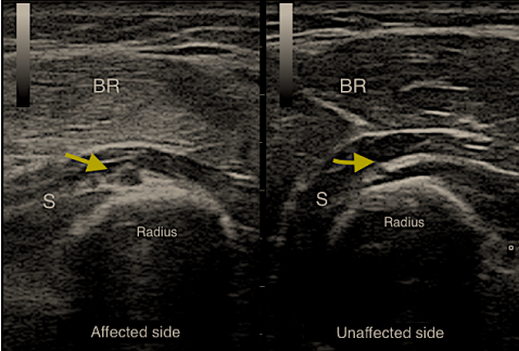
A 35-year-old man had fallen down from the first floor and admitted emergency department with pain, swelling and deformity on the right elbow. The radiograph showed fracture of the right supracondylar humerus. After surgical exploration and internal fixations, the patient referred to our clinic as elbow contracture with traumatic median and ulnar nerve injury. The surgeon noted that radial nerve was seen as intact. In physical examination strength of extensor carpi radialis, extensor digitorum communis, extensor carpi ulnaris and lumbrical muscle grades were about 1/5. We also observed a sensory deficit in the radial nerve dermatome. In ultrasonographic examination, we observed thickening in the nerve, increased hypoechogenicity and loss of neuronal fascicle distinction at the distal part of the elbow. No power Doppler signal was observed as a sign of neovascularization (Figure 1). Electrodiagnostic studies also showed severe axonal injury to the radial nerve. Motor study of the radial nerve showed decrease in the compound muscle action potential (CMAP). In addition, needle electromyography examinations of the muscles innervated by the radial nerve were consistent with acute denervation.
 Figure 1: The yellow arrow shows radial nerve both affected and unaffected side. On the affected side thickening in the nerve, increased hypoechogenicity and loss of neuronal fascicle distinction at the distal part of the elbow was observed.
BR: Brachioradialis muscle; S: Supinator muscle. View Figure 1
Six weeks after the rehabilitation programme he regained the ability to extend the fingers but he was still unable to extend the wrist. Because of this improvement conservative management was continued. After three months, the control electrodiagnostic studies showed decrease in the abnormal spontaneous activities and reinnervation findings were observed. Wrist extension movement was also started.
The radial nerve is the most injured peripheral nerve after long-bone fractures [1]. Especially supracondylar humerus fractures are commonly accompanied by nerve injuries due to closed proximity between radial, median and ulnar nerves. The nerves may be injured as a result of initial trauma or during fracture reduction. Surgical exploration for traumatic neuropathies is an effective method for evaluation [2]. In this report we presented a case with misdiagnosed radial nerve injury after supracondylar humerus fracture and internal fixation.
Most of the reports about radial nerve injury used the electrodiagnostic studies to evaluate the nerve damage [3,4]. On the other hand ultrasonography has become a widely used method to evaluate peripheral nerve palsies. Bodner, et al. showed that ultrasonography can be performed after humeral fractures to evaluate the radial nerve [5]. It seems to be advantageous because it is more comfortable for the patient and requires shorter time than electrodiagnostic studies. Physicians may use this useful tool as well as in follow-up for nerve damage recovery.
Authors declare that they have no conflicts of interest or any financial support.