It is about a 68-year-old woman who presented to the emergency department with a painful inflammatory swelling in the right upper abdomen and the epigastrium since 6 days. She had a medical history of obesity and type 2 diabetes. She had no surgical history. At physical examination, there is no fever with tenderness in the right upper quadrant of the abdomen. She presented a lump in the right upper abdomen and the epigastrium measured 5 × 6 cm which is red and painful. Laboratory data showed white blood cells at 14000/mm3, C-reactive protein at 160 mg/l. The computed tomography of the abdomen revealed (Figure 1) a large peripherally enhancing fluid-filled collection within the subcutaneous tissue of the right upper anterior abdominal wall, measuring 5 × 6 cm. The collection extended into the abdominal cavity, with a contiguous collection adjacent the region of the gallbladder fossa. The gallbladder was distended with thickened wall. Cholecystectomy with abscess drainage was performed. The postoperative course was uneventful.
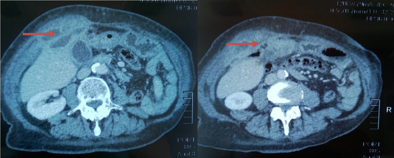 Figure 1: Computed tomography of the abdomen showing collection in the subcutaneous tissue of the anterior abdominal wall extending into the abdominal cavity (red arrow).
View Figure 1
Figure 1: Computed tomography of the abdomen showing collection in the subcutaneous tissue of the anterior abdominal wall extending into the abdominal cavity (red arrow).
View Figure 1
This is an unusual case. It is exceptional for cholecystitis contents to drain from an anterior abdominal wall abscess [1]. Such complication may result from eventual perforation of the gallbladder [2]. This complication is generally observed in chronic cholecystitis.