Retained surgical sponges (gossypiboma) are very rare but occur even under the presumed correct sponge counts postoperatively. Gossypiboma has the ability to cause significant harm to the patient and carry heartfelt medicolegal and professional repercussions to clinicians and hospitals. Its mani-festations may be non-specific and may take weeks, months or even years from the time of surgery. Therefore, diagnosis is based on a high index of suspicion with careful assessment of the patient's history, physical examination, and investigation. Retained sponges may extrude externally through a fistulous tract or internally into the rectum, vagina, bladder, intestinal lumen or through direct migration; however, intra-luminal migration is relatively rare. It is in consonance with this rarity that we report a case of an intestinal obstruction secondary to an intra-luminal foreign body. Intra-operative findings revealed a laparotomy towel accidentally left behind during a laparotomy for a ruptured acute appendicitis.
Intestinal obstruction, Laparotomy, Gossypiboma, Laparotomy towel
Gossypiboma is a term used to designate a mass of cotton material, commonly, sponges, gauze and towels, unwittingly left in the body at the end of surgical operation. This condition is called gossypiboma is derived from Gossypium (cotton in Latin) [1] and boma (place of concealment) [2]. It is one of the principal causes of intestinal obstruction with unknown incidence rate due to the fear of medico-legal implications. However, it has been reported as 1 in 100-3000 for all surgical interventions and 1 in 1000-1500 for intra-abdominal operations [3].
The most significant causes are emergency surgery, sponge retention, unplanned change in operation and high body mass index while disorganization, hurried sponge counts, long operations, unstable patient condition, inexperienced staff, inadequate staff numbers are less significant causes [4]. Retained sponges are most frequently observed in patients with obesity, presenting with non-specific abdominal pain and intestinal obstruction. Intramural migration of a sponge may occur without any opening to show its point of entry during re-exploration [5,6]. Inside the bowel lumen peristalsis pushes it on. If it can negotiate the ileo-caecal valve it will be passed out during defecation; if not it may cause intestinal obstruction, malabsorption or hemorrhage [7,8].
The diagnosis may be difficult and may mimic a benign or malignant soft tissue tumour in the abdomen and pelvis because symptoms usually are non-specific and may appear years after the antecedent surgery [9]. Radiological diagnosis can be made by ultrasonography showing an echogenic lesion with a hypoechoic rim and strong posterior acoustic shadowing whilst in some studies, plain X-rays of the abdomen reveal a long and fine opacity which is difficult to appreciate due to lack of radio-opaque impregnation of surgical sponges. A whorl-like spongiform hypodense mass with a thick peripheral rim on CT scan is the most common finding. On MRI, a retained sponge is typically seen as a soft tissue density mass with a thick well-defined capsule [7]. Although there are no known measures to completely eliminate the risk, preventive strategies aim primarily to increase awareness.
A 62-year-old patient presented to the accident and emergency unit of Komfo Anokye Teaching Hospital (KATH) with complaints of colicky dull abdominal pains, constipation, vomiting and gradual progressive abdominal distension for three weeks but passed flatus. She has a long history of diabetes and hypertension. Abdominal pain was non-radiating with no relieving and aggravating factors. Patient had laparotomy with appendicectomy done 17 months ago on account of histopathologically confirmed ruptured appendicitis. She has been reporting subsequently with non-specific abdominal pain until worsening of her current condition.
On general examination, she was stable, obese with a body mass index (BMI) of 30, temperature 35.6 ℃, not pale, anicteric, satisfactory hydration and a pulse 112 bpm RGV. As regards to her vital signs, her blood pressure (BP) was 142/89 mmHg, respiratory rate of 18cpm and oxygen saturation 98% on room air. She had urine output of 86 ml/hr.
Local examination revealed abdominal distention, with a presence of a lower midline incision scar, generalized tenderness, guarding; however, intact hernia orifices were intact. Plain abdominal radiographs showed multiple air-fluid levels and an abdominopelvic ultrasound suggested no intra-abdominal mass, free fluid or lymph nodes noted, while the bowel loops of normal wall thickness with normal peristaltic activity.
Pre-operative diagnosis of intestinal obstruction secondary to postoperative adhesions was made and patient was optimized for surgery and intravenous antibiotics started. She underwent laparotomy; the abdomen was explored through a median incision revealing multiple adhesions between bowel loops and a palpable mass in the sigmoid colon. Bowel proximal to mass was dilated and rectum was collapsed. The sigmoid colon (Figure 1) was resected and end-to-end colorectal anastomosis done to restore bowel continuity. It was noticed that the cause of the obstruction was a migrated sponge into the lumen. The post-operative period was uneventful, and patient was discharged on the 10th postoperative day. She was subsequently reviewed on two occasions after discharge and had no complaints.
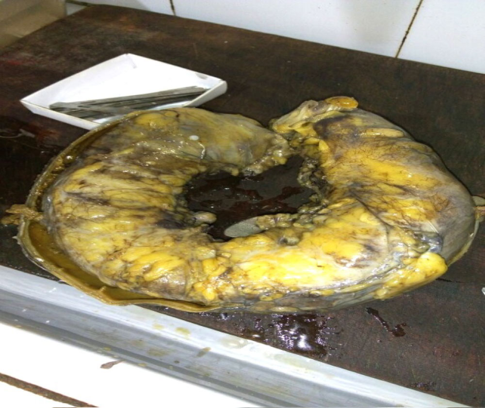 Figure 1: Resected sigmoid colon with a palpable mass. View Figure 1
Sigmoid colon was then dissected by a pathologist (Figure 2) when sample was sent for histopathology, revealing a 15 × 10 cm laparotomy towel (Figure 3).
 Figure 2: Dissection of sigmoid colon exposing a foreign body. View Figure 2
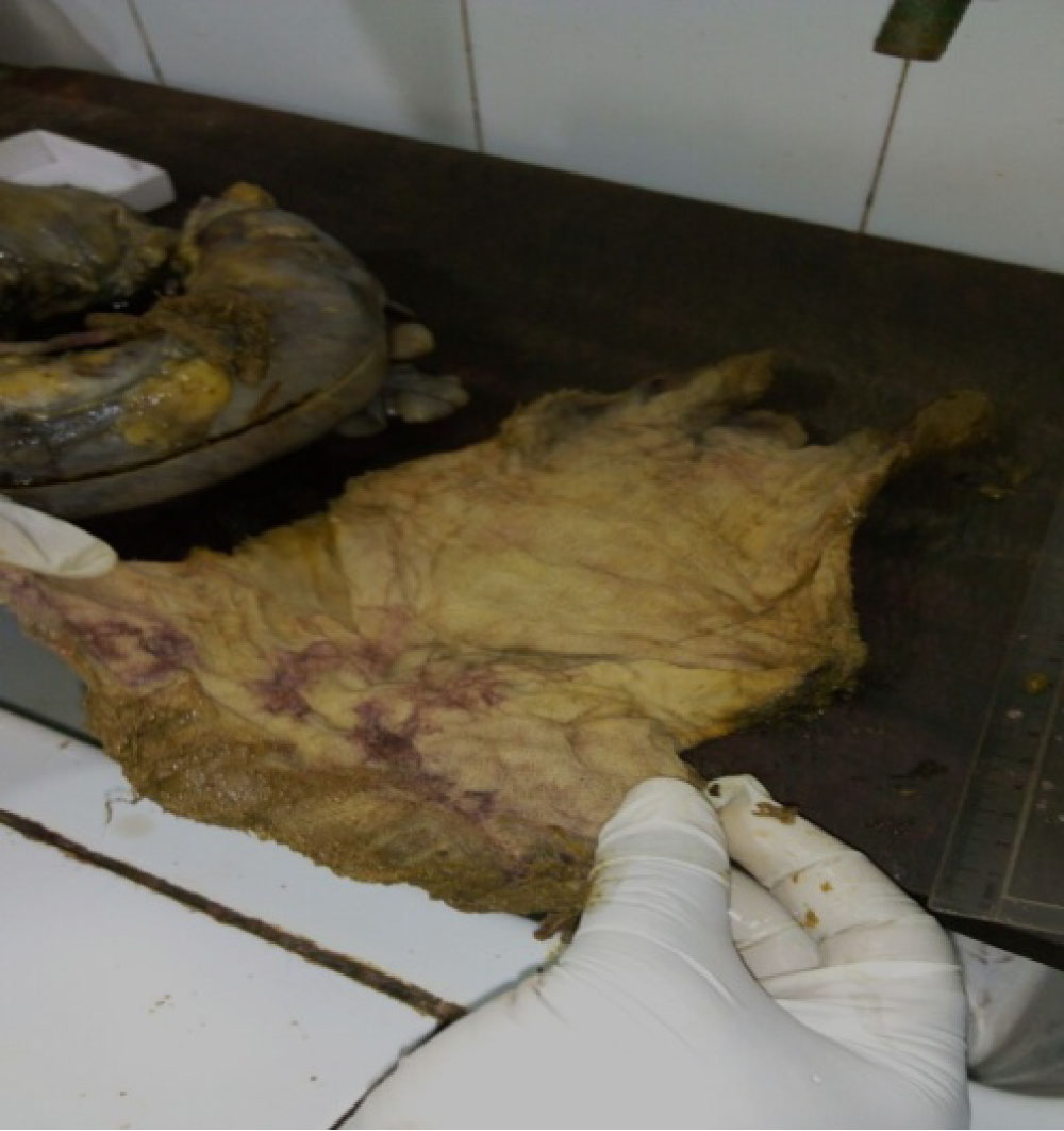 Figure 3: 15 × 10 cm laparotomy towel found in the sigmoid colon. View Figure 3
A gossypiboma may lead to foreign body reactions of two type's i.e, formation of foreign body granuloma due to aseptic fibrinous response or exudative reaction leading to abscess formation [10]. Migration of retained sponge into bowel is rare compared to abscess formation and occurs as a result of inflammation in the intestinal wall that evolves to necrosis [11]. Then the intestinal loop closes after complete migration of sponge. The defect in the bowel wall is sealed off by the wall of the granuloma and this prevents discharge of intestinal content into the cavity [12]. In the presented case, a similar process might have taken place. The retained sponge may be extruded along the large bowel and out of the anus or get lodged in the large bowel or at the terminal ileum causing an intestinal obstruction as happened in the case presented above [13].
Average discovery time in a study conducted by Sumer et al equaled 6.9 years with a median of 2.2 years [14] however, in our patient; the gossypiboma was detected at 1.4 years from the antecedent surgery. Patient's presentation was suggestive of a retained foreign body as a result of initial laparotomy for a ruptured appendicitis. The symptoms of gossypiboma depend upon the size of the swab, location and body reactions that manifests. It may present early with pain or may remain asymptomatic for long time with indeterminate symptoms. Moreover, patients may also present with subacute intestinal obstruction or abdominal mass [15]. In our case, she kept visiting the facility with non-specific abdominal pain which progressively worsened with later obstructive symptoms. Plain abdominal radiographs confirmed intestinal obstruction, but the abdominal ultrasound was unable to detect retained sponge. Risk factors identified in the case presentation for gossypiboma were obesity (BMI of 30), sponge count performed just once, length of surgery was one hour two mins and it was emergency surgery. The patient eventually underwent an exploratory laparotomy with resection of sigmoid mass (gossypiboma) and end-to-end anastomosis. Nosher and Siegel described 6 patients in whom percutaneous extraction was also successfully performed for removal of foreign bodies including an intra-abdominal laparotomy towel, 2 pelvic drains, and an angiographic guide wire fragment and a superficially located sewing needle and bullet [16].
Foreign bodies inadvertently retained in the abdominal cavity range from small gauzes and sponges to surgical instruments. Retention of these in the abdominal cavity is uncommon because it is under-reported and can carry serious medico-legal consequences [17-19]. These instruments are usually inert and can cause intestinal obstruction if they compress a section of bowel or the bowel is caught in the jaws of the instrument or lodged intra-luminally getting obstructed at the terminal ileum or large intestines [12,20].
Surgeons should be acutely aware of the risk factors that result in retained sponges and make deliberate efforts to prevent its occurrence. Interventions such as keeping a thorough pack and instrument count once at the start and twice at end of surgery, tagging the packs with radio-opaque markers, performing methodical wound examination and abdominal exploration before closure, avoidance of small sponges during laparotomy, avoidance of staff changes during procedures and in the event of doubt of accuracy of final count, intra-operative radiologic screening or re-exploration must be done. A high index of suspicion is required to diagnose gossypiboma and prompt commencement of intervention to reduce patient morbidity and mortality [5,21].
Gossypiboma is asymptomatic and weighty postoperative complication. It has grave implications for the patients and the surgeon. Gossypiboma can be avoided and can be achieved when surgeons follow preventive strategies such as regular pre-and post-operative sponge counts and use of radiopaque markers. Late presentation of gossypiboma can be far-reaching diagnostic conundrum and therefore proper examination should be conducted postoperatively for patients with atypical symptoms and a history of prior surgery.