Fibrosing Mediastinitis (FM) is a rare but increasingly recognized complication that has been mainly associated with chronic pulmonary histoplasmosis, granulomatous diseases, infections and autoimmune processes. Symptoms vary depending on compromised structures, mainly structures within the mediastinum. This review will focus on the etiology, clinical presentation and management of fibrosing mediastinitis based on multiple case series published to date.
1. Fibrosing mediastinitis is a rare, mainly associated with histoplasmosis infections. Also associated with sarcoidosis, tuberculosis, malignancies as well as other autoimmune processes.
2. Hallmark features include fibrosing sclerosis encompassing and compromising mediastinal structures, including inferior and superior vena cava, pulmonary artery and vein, esophagus and heart.
3. Treatment approach depends on symptomology, structures involved and include medical management, surgical management and conservative/palliative options.
Fibrosing Mediastinitis (FM) is a rare complication that has mainly been associated with histoplasmosis infections, however it can also be a complication of granulomatous infiltrative processes, including tuberculosis and sarcoidosis. The results of this condition can lead to significant symptom burden that require a unique approach to its management. We submit a comprehensive review concerning the natural history and management of fibrosing mediastinitis.
Fibrosing mediastinitis, Pulmonary histoplasmosis, Granuloma, Mediastinum, Histoplasmosis capsulatum, Fibrosis
Database searches conducted during the initial review done via institutional database proxy. Access granted through shared agreement with medical education and partnering medical institution. The database search was conducted using the key terms "fibrosing mediastinitis" and "fibrosing plus histoplasmosis". These terms were chosen due to the specific nature and etiology of the presenting case. During this initial database search, over 600 articles identified. Further truncating done to eliminate articles not within the last 30 years. Exception made for early case studies [1]. All articles cited were fully accessible electronically. Fifty total articles were screened with 28 articles directly cited in the preceding work. Further articles were eliminated due to relevancy (Figure 1).
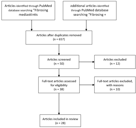 Figure 1: Fibrosing Mediastinitis. View Figure 1
Fibrosing mediastinitis, although not well understood, is a sequalae or progression of an underlying infection or granulomatous disease. It has been highly associated with histoplasmosis especially in the United States (US) [2]. It has been described in case reports associated with other disease processes, including infectious causes such as blastomycosis, sarcoidosis, tuberculosis aspergillosis, mucormycosis and cryptococcosis [3-5]. Some autoimmune causes have also been reported, including Bechet disease, rheumatoid arthritis and systemic lupus erythematosus [5]. Other associated causes include adenocarcinoma of lung, Hodgkin's disease, and associated treatment with radiation therapy [5-7] (Table 1). There have been many postulated mechanisms including an abnormal immunological response to the infection or autoimmune process [2]. It is thought that during this response, acellular collagen and fibrous tissue overwhelm the mediastinum, causing severe obstruction, compression and compromise of mediastinal structures [8].
Table 1: Causes of Fibrosing Mediastinitis. View Table 1
Most case reports are isolated to those geographical regions endemic for histoplasmosis, specifically the Midwest region [9]. Epidemiological studies have identified Ohio, Indiana and Arkansas as the states with most incidences of H. capsulatum based on Medicare claims data from 1998-2009 [10]. Exact epidemiological information is difficult to assess, as majority of patients infected with H. capsulatum are asymptomatic. Outside the US, most cases are unreported, however case series have been reported in the United Kingdom [5].
To date, two large case series identifying 80 and 94 patients with suspected FM has been reported [11,12]. Both were single center retrospective reviews. Of note, the Mayo Clinic electronic chart review found 54% of patients were female, and found the average age in years to be 42 [11]. In that clinical review over a 10-year period (1998-2007), age ranged between 21-75 years of age. In a previous clinical case review from 1975-1984, Garrett, et al. identified the average age of 33 years of age, with a range from 1-83 years old with the peak age between 20-40 years of age [12]. Similar sex distribution was noted as well. No race distinction was made in the Mayo clinic review by Peikert, et al. however Garrett noticed that majority of patients were Caucasian versus African American, 81% to 19% respectively. A single center study of twenty Asian patients over a 10-year period found predominantly female with and mean age of 69.5 years [13].
Clinical presentation varies dramatically. The structures involved the degree to which they are compromised and the duration in which they have been compromised will determine symptomology. Structures reported to be involved in fibrosis and subsequent obstruction include the pericardium, mainstem bronchus, esophagus, Superior Vena Cava (SVC), Pulmonary Artery (PA), Pulmonary Vein (PV) and phrenic nerve [14]. In the Peikert, et al. study, the mediastinal structures were compromised in 98% of patients, mainly the large airways, SVC and PA. It is important to delineate those who have significant pulmonary artery compression leading to pulmonary hypertension [15]. Seferain, et al. retrospectively identified 27 such patients who had pulmonary hypertension as a result of mediastinal fibrosis. Symptomology can vary from dyspnea, cough, and chest pain to frank hemoptysis due to airway invasion or great vessel obstruction [1]. The nature of fibrosis can lead to varying degrees of complications, with one report attributed mediastinal disease and chylothraces [16]. Another case report reported right carotid artery stenosis in a female patient [17]. Other associated complications include SVC syndrome and esophageal compression [11] (Table 2).
Table 2: Comparison of symptoms across case series. View Table 2
Due to its association with H. capsulatum, most patients usually have a diagnosis via serological assay or bronchioalveolar lavage [11,18]. Definitive diagnosis for histoplasmosis has been achieved via antibody titers or Grocott methenamine silver stains revealing H. capsulatum [11]. There is no definitive laboratory assay to diagnose FM. Due to its host immune modulated response, some genetic markers related to human leukocyte antigen markers has been identified, however more definitive studies are lacking [19]. Chest radiographs (Figure 2) can assist in diagnosis, usually showing enlarged perihilar and mediastinal lymph nodes. It is important to note that some chest radiographic findings are non-specific, including widening of the mediastinum, cavitary lesions, and adenopathy, prompting further workup with chest Computerized Tomography (CT) and/or bronchoscopy [8] (Figure 3 and Figure 4). Chest CT usually shows focal or diffuse fibrosis, usually confined to the middle mediastinum [20]. The focal appearance of the fibrosis appears more frequently in cases of FM than the diffuse pattern. Rarely is the posterior mediastinum involved [21]. As with most cases of FM, contrast enhanced imaging will depend on the structures involved. If vasculature is involved, contrast enhanced CT will benefit in confirming the diagnosis (Figure 5 and Figure 6). Other modalities, including MRI, esophagram or contrast pulmonary arteriogram may be useful to rule out malignancy or other mass effect causing lesions. It is important to note that the other modalities are not as efficient in representing the degree of calcification and fibrosis, therefore CT imaging should be the modality of choice if FM is suspected [8,20,22].
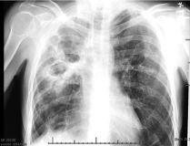 Figure 2: Initial Right Upper Lobe (RUL) cavitary lesion seen on PA Chest X-ray. View Figure 2
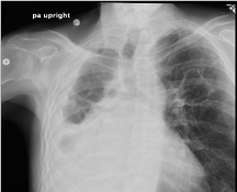 Figure 3: Disease progression seen on PA chest X-ray with tracheal deviation. View Figure 3
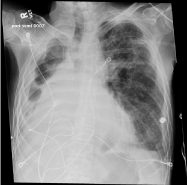 Figure 4: PA chest x-ray showing complete destruction of right lung and progressive fibrosis of left lung. View Figure 4
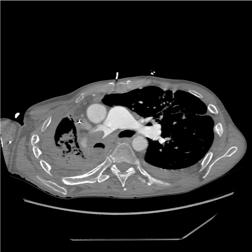 Figure 5: CTA Chest Contrast showing encroachment of main pulmonary artery by disease. View Figure 5
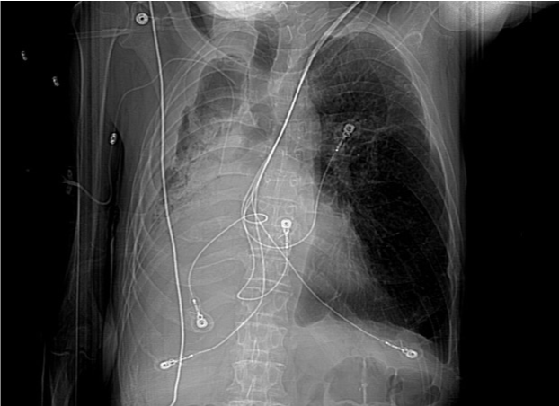 Figure 6: AP View chest with tracheal deviation and complete destruction of right lung. View Figure 6
The role of bronchoscopy in diagnosing fibrosing mediastinitis is limited to assisting in exclusion of other pathology. Diagnosis of H. capsulatum can be done via Bronchioalveolar Lavage (BAL). Other infectious etiology can be ruled out as well. Once case series of three patients reported that bronchoscopy was inconclusive in diagnosing FM, and findings closely mimicked that of chronic bronchitis [23], with bronchial narrowing, hyperemia and mucosal edema.
In the largest clinical case series reported to date, biopsy was not pursued routinely [11]. Surgical resection was done for pathology, with findings consistent with fibrosis and granulomatous inflammation. It is important to note there is no specific histological criteria for diagnosing FM.
It must be noted that treatment options are not curative. There is only symptomatic management, ranging from medical, surgical, interventional radiologic to palliative measures.
Management selection is based on symptom burden, patient comorbidities and ability to tolerate medical interventions.
Medical treatment options vary in opinion, practice and outcome. Ranging from antifungal treatment to corticosteroid treatment [24]. Treatment is aimed at controlling the underlying disease process leading to the fibrosis, and not the fibrosis itself. In one case series of 71 patients, treatment with amphotericin was not recommended, and showed no benefit in outcomes [1]. Corticosteroid treatment was also not recommended routinely in treating fibrosing mediastinitis as it does not alter the progression of symptoms. It was noted in the same case series, however that if there is bronchial hyperreactivity leading to wheezing or periodic dyspnea, then corticosteroids can be considered on a case by case basis [1]. Another case review of 80 patients at a single center revealed 34 patients were recommended for medical therapy, mainly antifungal or anti-inflammatory treatment (i.e. prednisone or tamoxifen) [11]. More recent guidelines from the IDSA do not suggest routine treatment with antifungals for fibrosing mediastinitis [25]. They do however suggest that 12-week course of anti-fungal therapy (itraconazole 200 mg once to twice daily) may be beneficial if radiographic findings are not conclusive for mediastinal fibrosis, however evidence of this practice is limited [25]. The thought process is that if there is still an acute inflammatory response, evidenced by elevated ESR, clinical findings suggesting acute inflammation, anti-fungal therapy may be beneficial as compared to chronic fibrotic sequelae [25]. As evidenced, medical treatment options are limited, and little is known about whether survival is affected by routine administration. In the latter study, no patient achieved complete remission from disease, and only 18% of those who followed up had any evidence of radiologic or symptomatic response to treatment [11].
Surgical options are available, and depend greatly on anatomical location of obstructions, fistulas, and most importantly symptomology and disease burden. Of note, unilateral disease is often more amendable to surgical intervention, and/or observation [14]. As with any surgical approach, careful consideration should be made to assess the patient's ability to withstand such a procedure and its associated high mortality. One single center study of 9 patients with mediastinal fibrosis, three patients underwent pneumonectomy [14]. Complications included bleeding, cardiac tamponade, and bronchial esophageal fistula. Other options for treating fibrosing mediastinitis include intravascular or pulmonary bronchial stenting [11]. One recent study of only four patients identified percutaneous pulmonary or artery vein stenting as palliative measure [26]. Although limited in size of study, outcome was generally favorable in these patients. Interventional procedures are also available, including embolization of offended vessels, including Superior Vena Cava (SVC), pulmonary artery or pulmonary vein. These procedures, often technically challenging are not viewed as favorably, according to literature review [11]. A surgeon or interventional radiologist who is proficient in performing such procedures, with the awareness of complications is necessary. One study identified 2 cases that required unscheduled open procedures after failed bronchial stents or bronchoplasty [11]. The evidence of fibrosis seen during mediastinoscopy or thoracotomy will determine the outcome of surgical release of offended structures. One study noted that surgical intervention at that point is usually technically impossible, hazardous and nearly fatal [12].
Most patients who suffer from symptomatic FM are able to return to a functional performance status. Reported cases of fatal FM are related to severe bilateral disease, and significant vascular or airway compression [11,27]. Some surgical methods can be deemed as palliative especially when structures are being compromised, including airway or esophagus. If surgical options are deemed unsafe with sure mortality, palliative options should be considered to relieve symptoms, including dyspnea associated with hypoxia, chest pain and anxiety. Not all patients with fibrosing mediastinitis have sure fatal outcomes [28,29]. Those who present asymptomatically tend to have better prognosis [11]. In the largest retrospective study to date, Piekert, et al. noticed a 100% survival rate 7 years after diagnosis, and greater than 95% survival rate after 14 years. Those patients who died, had bilateral disease likely related to disease extension [11]. Advances in disease reporting and novel therapeutic interventions are likely contributing to increased survival.
Although rare, fibrosing mediastinitis is an insidious complication of pulmonary histoplasmosis that must be recognized quickly, along with rapid stabilization and carefully crafted treatment plan. Medical therapy with antifungals and corticosteroids is not recommended routinely. Surgical intervention should be considered carefully, including approach and clinical outcome. Palliative measures should be considered especially in those with increased disease burden and structural involvement. More defined diagnostic criteria and treatment algorithms are needed to assist in recognizing and treating FM.