IMAGE ARTICLE | VOLUME 4, ISSUE 4 | OPEN ACCESS DOI: 10.23937/2474-3682/1510097

A Rare Case Report: Cystic Spinal Meningioma

Serdar Arslan , Hasan Erdogan, Fatma Zeynep Arslan, Mehmet Sedat Durmaz, Fatih Oncu and Ismet Tolu

Department of Radiology, Konya Education and Research Hospital, University of Health Sciences, Konya, Turkey

*Corresponding author: Serdar Arslan, Department of Radiology, Konya Education and Research Hospital, University of Health Sciences, 42090, Meram, Konya, Turkey, Tel: +90-555-866-0621, E-mail: arslanserdar10@gmail.com

Accepted: July 24, 2018 | Published: July 26, 2018

Citation: Arslan S, Erdogan H, Arslan FZ, Durmaz MS, Oncu F, et al. (2018) A Rare Case Report: Cystic Spinal Meningioma. Clin Med Img Lib 4:097. doi.org/10.23937/2474-3682/1510097

Copyright: © 2018 Arslan S, et al. This is an open-access content distributed under the terms of the Creative Commons Attribution License, which permits unrestricted use, distribution, and reproduction in any medium, provided the original author and source are credited.

Keywords


Spinal, Cystic meningioma, MRI

A 41-year-old woman was admitted to our department presented with a 1-year history of persistent weakness and progressive numbness in the left lower limb. MRI revealed that there was a well-circumscribed intraspinal extradural lesion at L1 and L2 vertebral levels (Figure 1). The lesion was isointense on T1- and T2-weighted images and it was shown strongly enhancing except a small nodular cystic component in superior part of the lesion (Figure 2). The patient was operated and histopathological studies confirmed the mass to be a meningioma.

Cystic spinal meningioma is a rare form of spinal meningiomas. They are easily confused with other tumors with cyst formation and have been seldom reported in the literature [1-3]. Spinal meningiomas are common extra-axial solid lesions with distinguishing features, and most of them have a thoracic localization [1]. The lesions tend to be isointense on T1- and T2-weighted images, enhancing strongly after administration of contrast agent. Cystic spinal meningiomas with a typical features complicate the imaging work up and cannot be easily identified on MRI due to their rarity [3]. This type of clinical entity should be considered in the differential diagnosis of intraspinal extradural neoplasms.

Source(s) of Support


None.

Conflicting Interest


None.

Figures


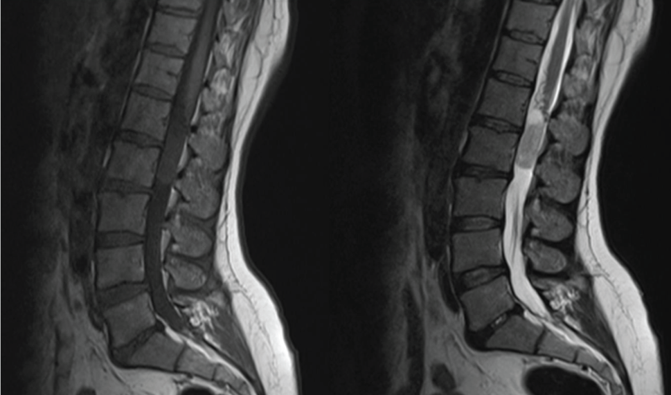
Figure 1: T1 and T2 weighted images show isointense well-circumscribed intraspinal extradural lesion. There is a small nodular cystic component in superior part of the lesion.


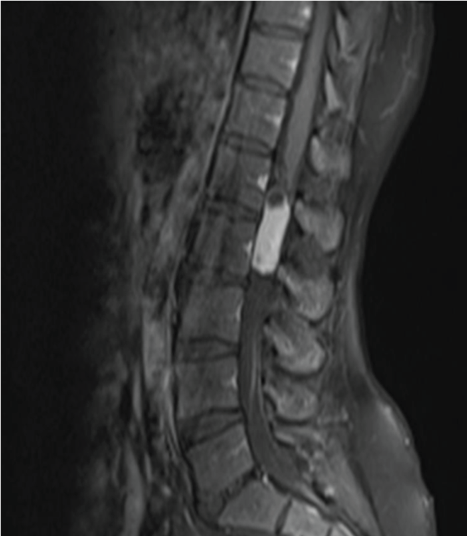
Figure 2: Intravenous contrast injection with fat sat T1 weighted image shows strongly enhancement of the lesion except a small nodular cystic component in superior part.

References


  1. EM Chatzidakis, Z Volikas, N Condilis, S Lypiridis, M Fratzoglou, et al. (2008) Spinal cystic meningioma presenting as a ring enhancing lesion on MRI. Ann Ital Chir 79: 197-198.

  2. C Sotiriadis, Q D Vo, R Ciarpaglini, HM Hoogewoud (2015) Cystic meningioma: Diagnostic difficulties and utility of MRI in diagnosis and management. BMJ Case Rep 2015.

  3. J Zhang, ZH Chen, ZF Wang, P Sun, JT Jin, et al. (2016) Epidural cystic spinal meningioma: A case report. Medicine (Baltimore) 95: e3030.