IMAGE ARTICLE | VOLUME 4, ISSUE 3 | OPEN ACCESS DOI: 10.23937/2474-3682/1510090

A Rare Case: Intrabiliary Obstruction due to Ruptured Hepatic Hydatid Cyst

Serdar Arslan , Hasan Erdogan, Fatma Zeynep Arslan, Mehmet Sedat Durmaz, Fatih Oncu and Ozgur Oner

Department of Radiology, Konya Education and Research Hospital, University of Health Sciences, Konya, Turkey

*Corresponding author: Serdar Arslan, MD, Specialist, Department of Radiology, Konya Education and Research Hospital, University of Health Sciences, 42090, Meram, Konya, Turkey, Tel: +90-555-866-0621, E-mail: arslanserdar10@gmail.com

Received: March 04, 2018 | Accepted: May 03, 2018 | Published: May 05, 2018

Citation: Arslan S, Erdogan H, Arslan FZ, Durmaz MS, Oncu F, et al. (2018) A Rare Case: Intrabiliary Obstruction due to Ruptured Hepatic Hydatid Cyst. Clin Med Img Lib 4:090. doi.org/10.23937/2474-3682/1510090

Copyright: © 2018 Arslan S, et al. This is an open-access content distributed under the terms of the Creative Commons Attribution License, which permits unrestricted use, distribution, and reproduction in any medium, provided the original author and source are credited.

Keywords


Hydatid cyst, Biliary, Ruptured, MRCP

A 69-year-old man was admitted to our department with a 2-day history of jaundice, nausea, and vomiting. MRCP showed that dilatation of the intra and extrahepatic bile ducts and fragmented membranes in the common biliary duct (Figure 1). Additionally, a hydatid cyst which is lost of volume tension was detected in segment 6 of liver (Figure 2). Impaction of hydatid material into the common bile duct was relieved endoscopically.

Cystic echinococcus is caused by the larval form of tape worm Echinococcus granulosus. Among the complications of hydatid liver disease, spontaneous cyst rupture into the biliary tract is unusual. Hydatid cyst rupture has been classified into three types: Contained (when only the endocyst ruptures and the cyst contents are confined within the pencyst); communicating (when the cyst contents escape via biliary radicles that have been incorporated in the pencyst); and direct (when both the endocyst and the pericyst tear, allowing cyst contents to spill into the pleural or pentoneal spaces) [1,2]. Most hydatid cysts of the liver eventually leak into small bile ducts or perforate into larger ones. Less frequently, a large bile duct is involved, allowing daughter vesicles and/or fragmented membranes to escape into the biliary tree. In this situation, obstructive jaundice or cholangitis is much more common than when the communication is small [3].

Source(s) of Support


None.

Conflicting Interest


None.

Figures


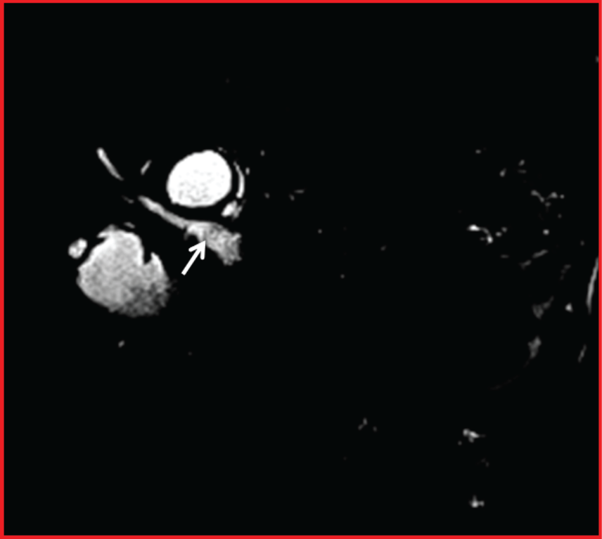
Figure 1: Magnetic Resonance Cholangiopancreatography (MRCP) shows dilatation of the extrahepatic bile ducts and fragmented membranes in the common biliary duct (white arrow).


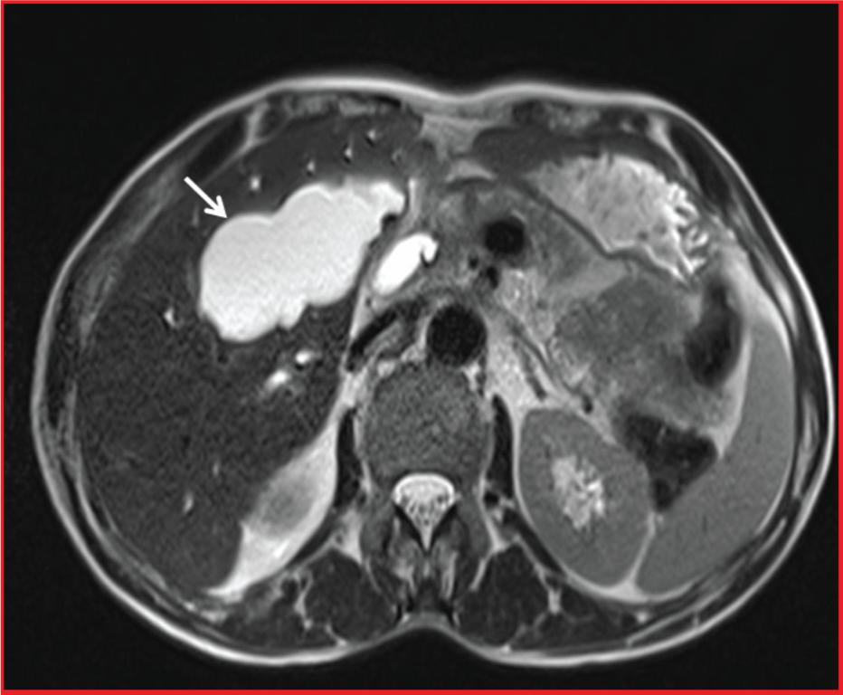
Figure 2: Axial T2 weighted image shows a hydatid cyst lesion with decreased volume and lobulated contour in the segment 6 of the liver (white arrow).

References


  1. Becker K, Frieling T, Saleh A, Haussinger D (1997) Resolution of hydatid liver cyst by spontaneous rupture into the biliary tract. J Hepatol 26: 1408-1412.

  2. Erden A, Ormeci N, Fitoz S, Erden I, Tanju S, et al. (2007) Intrabiliary rupture of hepatic hydatid cysts: Diagnostic accuracy of MR cholangiopancreatography. AJR Am J Roentgenol 189: 84-89.

  3. Arslan S, Bakdik S, Oncu F, Tolu I, Eryilmaz MA (2017) Successful percutaneous treatment of extrahepatic cystic echinococcosis through PAIR and single puncture catheter techniques. Jpn J Radiol 35: 296-302.