Calvarial hemangioma, Skull, CT, MRI
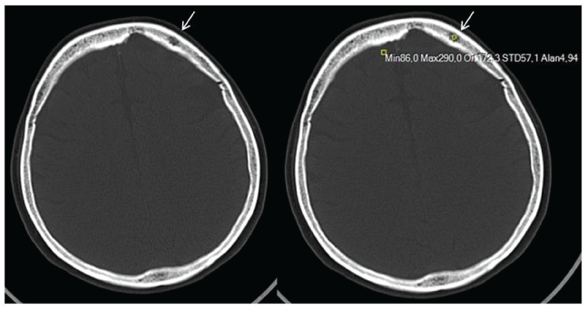
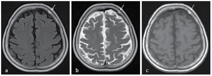
A 66-year-old female patient with a long time history of headache admitted to our hospital. There was no problem in laboratory findings. The patient underwent brain computed tomography (CT) and magnetic resonance imaging (MRI). Brain CT demonstrated a lytic, high density (mean 172 HU), characteristic trabecular thickening radiating from a common centre lesion at left frontal bone (Figure 1). MRI showed a smooth, lesion which was hyperintense in all sequences (Figure 2). The characteristic findings on CT and MRI suggested hemangioma of frontal bone.
Skull vault haemangiomas or haemangiomas of the calvaria, are benign slow growing vascular neoplasms affecting the skull diploe in any location [1]. These tumors, arising from the intrinsic vasculature of the bone, are mostly found in vertebral bodies and represent 0.2 percent of all benign neoplasms of the skull [2]. The majority of these lesions are asymptomatic, but patients can present with focal pain or a palpable mass [3]. The great majority of reported cases of hemangiomas are unifocal but multiple hemangiomas have been reported. Hemangiomas have been classified as cavernous which is predominant in hemangiomas of the skull, capillary or venous [3,4]. Plain radiography or CT scan reveals this lesion as solitary lytic lesion with a sclerotic rim, while MRI shows isointense on T1-weighted images and hyperintense on T2-weighted images, consistent with regions of slow-flowing blood [2,4]. Sometimes, the classic radiographic appearances are not evident and lesions can be misinterpreted as lesions like multiple myeloma or osteosarcoma. The gold standard treatment is en bloc resection of the tumor with the removal of a rim of normal bone [4].
None.
None.

Figure 1: Brain CT demonstrates a lytic, high density (mean 172 HU), characteristic trabecular thickening radiating from a common centre lesion at left frontal bone (arrows).

Figure 2: a) Axial T1 weighted; b) Axial T2 weighted; c) Axial FLAIR sequences showed a smooth, lesion which was hyperintense in all sequences (arrows).