Ludwig's Angina (LA) is a life-threatening emergency disease characterized with mouth floor and submandibular space cellulitis. LA frequently begins from submandibular region first and the tongue is pushed forward. Diabetic patients, immunocompromised conditions such as chronic hepatitis, chronic renal failure, chemotherapy are more prone to this condition. We are here to present the Computed Tomography (CT) and Magnetic Resonance Imaging (MRI) findings of a 41-year-old LA patient.
Ludwig's angina, Magnetic resonance imaging, Computed tomography
A 41-year-old male patient with diabetes mellitus was admitted to our hospital complaining of swelling in the mouth and fever. At the time of admission on laboratory finding; white blood cell was 27.21 IU/L (references: 4.0-10.0 X 10⁹/L), c-reactive protein was 252 mg/L (references: 0-5 mg/L), procalcitonin was 19.61 ng/ml (references: < 0.5 ng/ml) and increased.
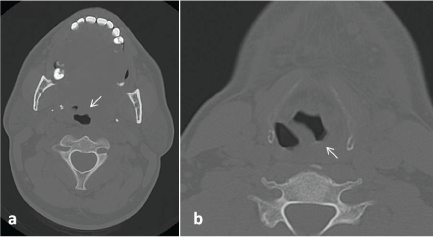
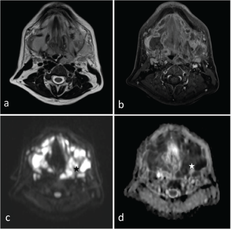
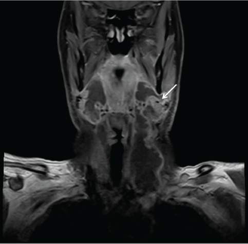
On USG; soft tissue thickening and tissue edema was seen in bilateral submandibular region, submental area and upper portion of the neck. Subsequently; CT was perfomed. CT revealed heterogeneous hypodense soft tissue thickening narrowing the air column in the left portion of the oropharynx at the level of the tongue base (Figure 1a). Left pyriform sinus is also seen as obstructed (Figure 1b). Neck MRI is performed with a clinical and radiological suspicion of LA and to be more confident about excluding a possible underlying malignancy. Neck MRI demonstrated subcutaneous soft tissue swelling and diffuse wall thickening of superior larynx, nasopharynx, oropharynx and hypopharynx (Figure 1). A cystic lesion with a 4.5 cm sized peripherally contrast enhancing was observed in the left peritonsillar area; diffusion restriction being demonstrated lesion was consistent with abcess (Figure 2 and Figure 3). In the submandibular and jugulodigastric areas, multiple LAPs were seen whose short axis was greater than 2 cm with a thickening cortex was observed. A significant clinical improvement was seen after iv amikacin and prednisolone initiation in patient who underwent surgical debridement. Cervical lymph node biopsies were reported as reactive hyperplasia on microscopic examination and surgically debrided tissue was pathologically confirmed as granulation tissue. Vancomycin and metronidazole were added to the antibiotherapy treatment and the patient was discharged with complete recovery.
LA was first described by Ludwig in 1836 as progressive gangrenous cellulitis and rapidly progressive and fatal infections of the soft tissues of the neck and floor of mouth [1]. One of the most frequent causes of LA is odontogenic-related [2]. The mostly encountered agents predominantly related to oral flora in LA are Streptococci, Staphylococci, Bacteroides and Fusobacterium spp. Before the invention of antibiotics LA was a mostly fatal infection; with the increased use of antibiotics fatality rates reduced but in the setting of spreading infection and edema via soft tissue may stil lead to end-stage airway obstruction, which could have devastating consequences [3]. Patients may have difficulty in breathing, swallowing. And trismus, fever and tenderness in the submandibular region, neck pain can be seen.
Diagnosis is mainly based on clinical findings; radiologically marked edema and diffuse wall thickening can be seen in the affected submandibular region, submental area and floor of the mouth especially in MRI. Due to its superiority in differentiating the cystic lesions from solid, USG may yield important information by demonstrating complications such as abscess. While MRI has certain advantages in terms of detecting soft tissues involvement compared to CT; both of these modalities are helpful in obtaining accurate diagnosis. Magnetic resonance angiography reveals vascular complications such as internal jugular venous thrombosis, carotid erosion or rupture more accurately [4]. It is important to keep the airway open in these patients; treatment also includes intravenous antibiotics, steroids and surgical debridement. In conclusion; LA is a life-threatening polymicrobial infection of the submandibular region. It is important for radiologists to be familiar with these rare appearances and to alert clinicians for early diagnosis.

Figure 1: a) At the level of the tongue base, heterogeneous hypodense soft tissue thickening narrowing the air column is seen in the left portion of the oropharynx (arrow); b) Left pyriform sinus is seen as obstructed (arrow).

Figure 2: a,b) Axial T1, T2 has demonstrated subcutaneous soft tissue swelling and diffuse wall thickening of superior larynx, left portion of nasopharynx, oropharynx and hypopharynx; c,d) On diffusion weighted axial sections have shown a cystic lesion with a 4.5 cm sized peripherally contrast enhancing is observed in the left peritonsillar area; Diffusion restriction being demonstrated lesion is consistent with abcess (star). In the submandibular and jugulodigastric areas, multiple LAPs were seen whose short axis was greater than 2 cm with a thickening cortex was observed.

Figure 3: On coronal plane; Diffuse soft tissue swelling and abcess in the left peritonsillar area is seen (arrow).