Duplication cyst, Gastric outlet obstruction, Adult, Endoscopy, Robotic surgery, Laparoscopy
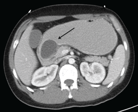
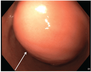
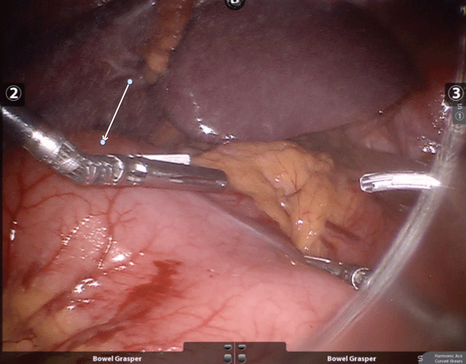
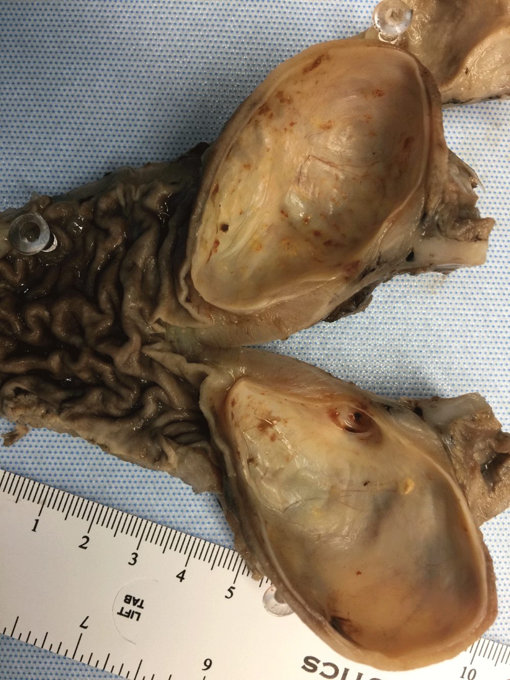
The patient is a 29-year-old man with no significant medical or surgical history who presented with 2 months of nausea, emesis, and 20 pounds of unintentional weight loss. On initial presentation, he was afebrile with normal vital signs. His abdomen was soft, non-distended and non-tender. Initial laboratory parameters revealed a hypokalemic, hypochloremic metabolic alkalosis consistent with a clinical picture of gastric outlet obstruction. Computed Tomography (CT) of the abdomen and pelvis (Figure 1) revealed a 3.5 × 4.9 cm cyst proximal to the pylorus, suggestive of a gastric duplication cyst, causing a gastric outlet obstruction. Endoscopy demonstrated a large pre-pyloric mass without noted mucosal abnormality or luminal connection, likely responsible for the gastric outlet obstruction (Figure 2). Antral biopsies revealed mild chronic inflammation and mild foveolar hyperplastic changes. Given gastric outlet obstruction as well as imaging suggestive of a duplication cyst, the patient was taken to the operating room and underwent robotic assisted laparoscopic distal gastrectomy with Billroth 2 gastrojejunostomy for a suspected gastric duplication cyst (Figure 3). Final pathology revealed features consistent with a gastric duplication cyst (5.2 × 4.0 × 4.0 cm) not communicating with the gastric lumen (Figure 4).
Gastric duplication cysts, rarely diagnosed in adults, and often discovered incidentally on imaging, were first reported in 1911 [1]. They are congenital abnormalities, and account for 2-4% of gastrointestinal tract duplication cysts [2]. They may present with non-specific symptoms including pain, nausea, vomiting, and weight loss. CT, magnetic resonance imaging, and Endoscopic Ultrasound (EUS) may assist in the diagnosis. EUS guided biopsy may help when other diagnoses, such as gastrointestinal stromal tumors, are suspected. In the stomach, most duplication cysts arise along the greater curvature; they are rarely reported in the antrum or pre-pyloric region. Malignant degeneration is rare [3,4]. Treatment of gastric duplication cysts involves complete cyst resection and typically achieved with laparoscopic or open methods.

Figure 1: Computed Tomography (CT) of the abdomen/pelvis demonstrates a 3.5 × 4.9 cm cyst in the pre-pyloric antral area (arrow) with associated gastric dilation suggestive of gastric outlet obstruction.

Figure 2: Endoscopy revealed a submucosal mass without luminal communication (arrow) corresponding to the lesion noted on imaging.

Figure 3: Intraoperative photograph demonstrating the external appearance of the gastric duplication cyst (arrow).

Figure 4: Surgical pathology revealed features consistent with a gastric duplication cyst (5.2 × 4.0 × 4.0 cm) not communicating with the gastric lumen.