Computed tomography angiography, Radionuclide imaging, Aortic disease, Prosthesis, Inflammation
A 79 year-old man arrived at emergency department because of having abdominal pain for several months (almost one year). Abdominal pain was located in mesogastrio, it was late postpandrial, associated with paresthesias in the left lower limb and irradiated through both iliac fosses getting to the back.
Because of this pain he had been visiting the digestive department where they performed a gastroesophageal transit which reported a hiatus hernia without any other complications.
Visiting the digestive department for the last time he was performed a US study which revealed an 8 × 12.8 cm abdominal aortic aneurysm with extensive circumferential thrombosis.
He was asked for an aortic angio CT which concluded with the existence of an 8.3 cm abdominal aortic aneurysm so he was admitted to the hospital for monitoring and treatment.
Five days later he was treated surgically and a Zenith LP aortobiiliaca endoprosthesis was implanted.
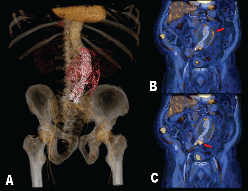
He was followed up during one year but fifty months later the angio CT revealed a juxtarenal abdominal aortic aneurysm excluded, with aortobiiliac endoprothesis, and a thrombosed aneurysm sac (Figure 1A) with a maximum size of 9 × 9.9 cm and true light of 25 mm; no signs of endoleaks or prosthesis related changes were observed but there was a paraaortic image to rule out inflammation so, a PET/CT was asked.
There was performed a 18F-FDG PET/CT study (Philips Gemini TOF) that showed not only a big infrarrenal abdominal aneurysm with areas of hypermetabolism on its wall (Figure 1B) but also hypermetabolic involvement in the distal part of the stent after the aortic bifurcation compatible with infection (Figure 1C).

Figure 1: A) PET/CT 3D reconstruction shows thrombosed aneurysm sac with aortobiiliac endoprothesis.
B) 18F-FDG PET/CT coronal view. Increased metabolic activity in the left side of the aneurysm sac wall (red arrow).
C) A focus with high glucose uptake was seen in the distal part of the stent after the aortic bifurcation compatible with infection (red arrow).