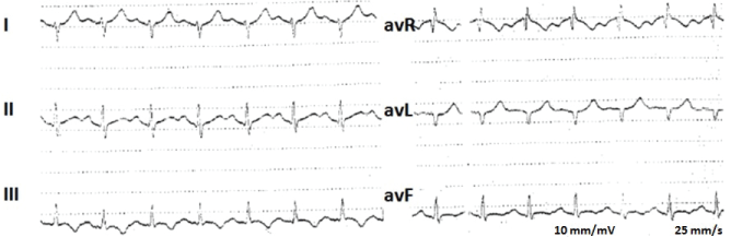
A 18 years-old woman presented to the Emergency Department with shortness of breath, pleuritic chest pain and syncope. She denied cough, sputum production, hemoptysis, wheezing, fever or leg pain. She had undergone an orthopedic surgery for comminuted fracture of the right lateral malleolus 15 days before this event. Her medical history was unremarkable. Her blood pressure was 126/80 mmHg; her oxygen saturation was 94% on room air. Chest auscultation was normal and revealed no adventitious sounds. A S1Q3T3 pattern and a sinus tachycardia were found in the electrocardiogram (Figure 1).
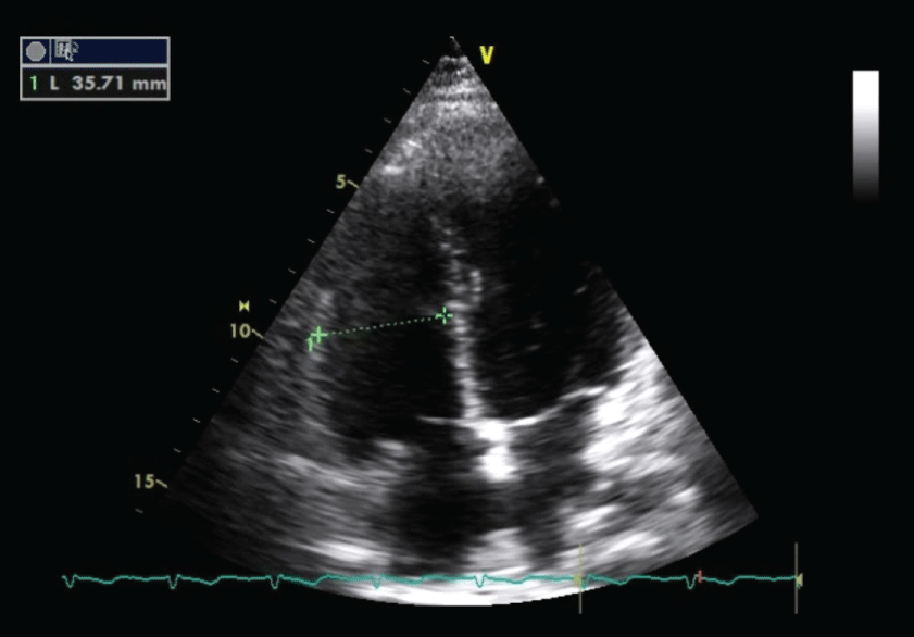
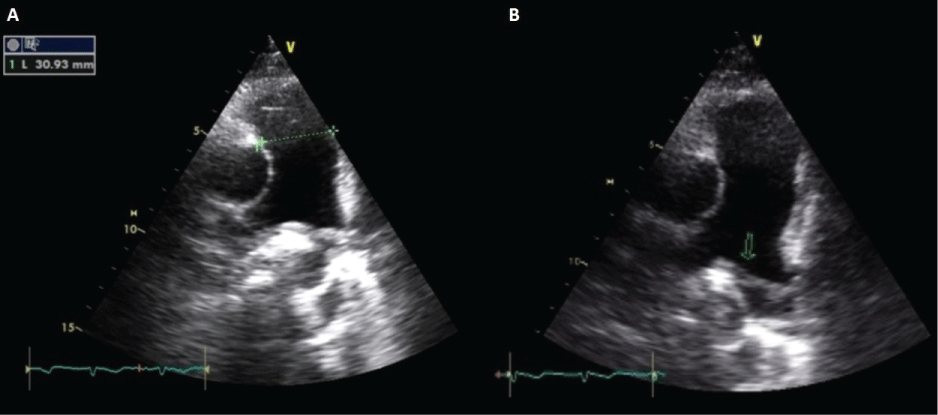
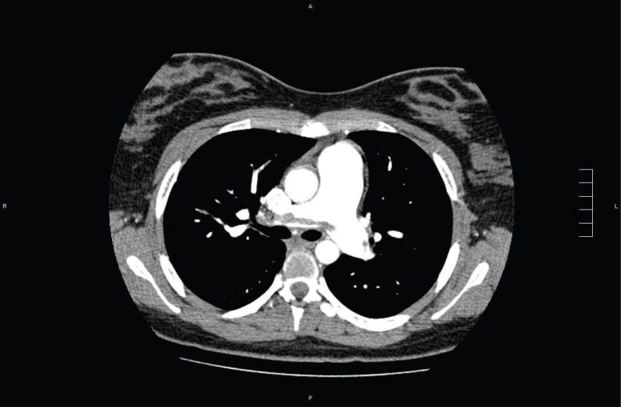
The focused echocardiogram showed slight right ventricle dilatation (Figure 2) and a slight enlargement of the pulmonary trunk and a thrombus in the pulmonary trunk bifurcation (Figure 3). Chest computed tomography with contrast showed multiple emboli up to the segmental level (Figure 4). Anticoagulation was initiated and the patient was admitted in the intensive care unit. She remained hemodynamically stable and she was discharged in a few days. One year later, the echocardiogram showed a normal heart.
Pulmonary embolism is a relatively common cardiovascular emergency. By occluding the pulmonary arterial bed it may lead to acute life-threatening, but potentially reversible, RV failure. Early diagnosis is fundamental. In this case, use of echocardiogram to evaluate the cause of the patient's critical state suggested the diagnosis. Increased familiarity with the use of bedside sonography in the evaluation of critically ill patient may allow clinicians to increase their efficiency in managing these patients.