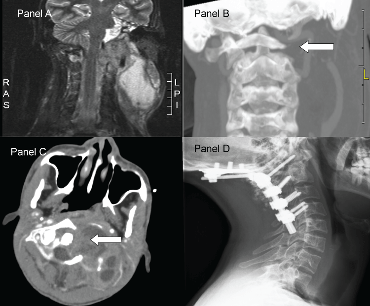
A 23 year old man was referred to the emergency room of our hospital because of a large swelling in de neck. The patient originated from Somalia and stayed in a centre for asylum seekers in The Netherlands for 6 months. His medical history told that he had a slow growing swelling of the left submandibular region, extending into the neck, which became painful and gave the patient discomfort and malaise. He also suffered chest pain on the right side. On clinical examination a large weak-elastic swelling was palpable in the neck on the left side, extending from the mandible to the clavicle. Because of pain and tumour mass there was a restriction of his head position to the left side. There were no neurological signs. Intraoral, a submucosal mass was visible on the left orofaryngeal wall. A gadolinium enhanced MRI (Panel A) showed a large multilocular partially solid, partially cystic process in the neck extending from the skull base towards the thorax aperture, together with intraspinal extension. The tumour process encased the vertebral artery, displaced the carotid artery and destructed the left occipital condyle and lateral mass of C1 (Panel B and Panel C). Furthermore, the 10th rib (right side) showed an osteolytic lesion. Based on CT and MRI the differential diagnosis could be infectious disease, Morbus Kahler, leukemia or lymphoma. Laboratory findings were suggestive of infectious disease. A biopsy of the cystic mass showed "acute, a specific infection"; no malignancy". PCR assessment of the biopsy material detected a tuberculosis mycobacterium complex. Because of craniovertebral junction instability an occipitocervical fixation was performed (Panel D).
In conclusion: On basis of the high prevalence of tuberculosis in Africa, patients from the African continent with single or multiple tumorous processes, in this case a single one in the cervical region together with an osteolytic lesion in a rib, should be diagnosed as having tuberculosis until proven otherwise.

Figure 1: