A 75-year-old female patient with past medical history of severe Alzheimer's disease, totally dependent with nasogastric tube feeding (NGT) presented to the emergency department with worsening dyspnea in the past 48 hours. Physical examination revealed on auscultation decreased air entry over the right lung and dullness to percussion. Two days before admission the patient had the NGT changed and a chest X-ray to corroborate position that was checked by one of the junior doctors and discharged to the nursing home. A thoracic bedside ultrasound demonstrated right pleural effusion. A chest radiograph revealed malposition of the NGT in the right pleural space and hydropneumothorax, the NGT was immediately removed without complications and thoracic drainage was inserted obtaining 1200 ml of enteral nutrition (Figure 1, Figure 2, Figure 3 and Figure 4). Her hospital course was satisfactory, and the patient was discharged 7 days later.
Hydropneumothorax secondary to misplacement of a nasogastric feeding tube.
Introduction of nasogastric feeding tubes is usually blindly performed and is considered a safe, easy, non-expensive procedure in awake patients [1]. The rate of complications of a blind insertion technique varies from 0.3 to 15% [1] and is usually related to inadvertent insertion of nasogastric tubes into the trachea and distal airways. Severe aspiration pneumonia, hydrothorax, hemothorax, empyema and delayed pneumothorax have been described as complications [2-5].
Physical examination hardly predicts malposition of the tube and the traditional bedside techniques of gastric aspiration and insufflation test lack specificity and sensitivity and often give false reassurance that the NGT is properly positioned, however, chest X-ray after insertion of feeding tube is considered a gold standard confirmatory test [6], which prevents additional complications, although in this case was misinterpreted. Plain radiography and chest CT are sufficient for a proper diagnosis of this serious complication, although sometimes laryngoscopy can be used to visualize the localization of the tube. Thoracic drainage is the technique of choice, nevertheless sometimes an isolated drainage is not sufficient and placement of additional chest tubes can be required or even performing a bronchoscopy to aspirate the feeding contents [7,8].
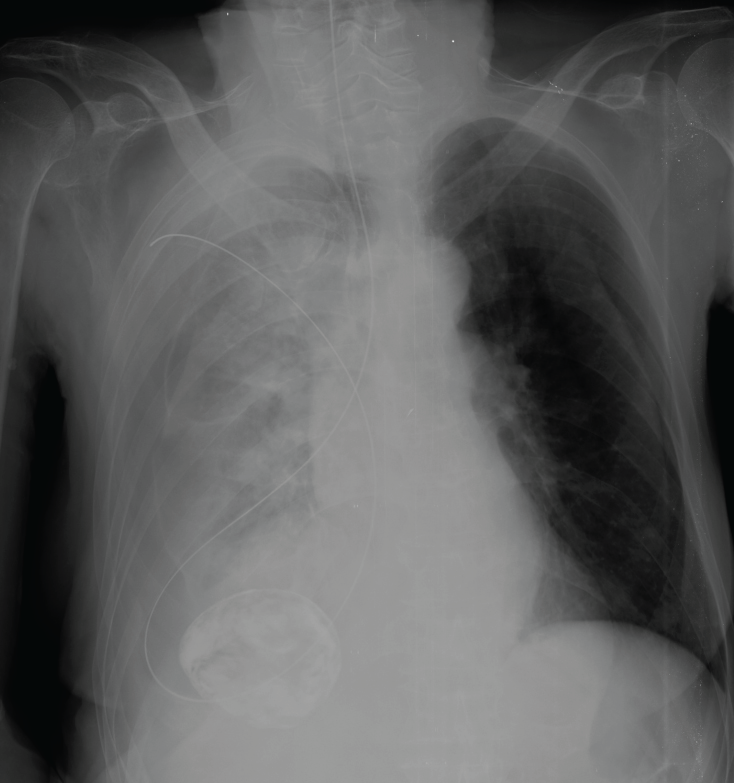
Figure 1: Nasogastric tube in the right hemithorax.
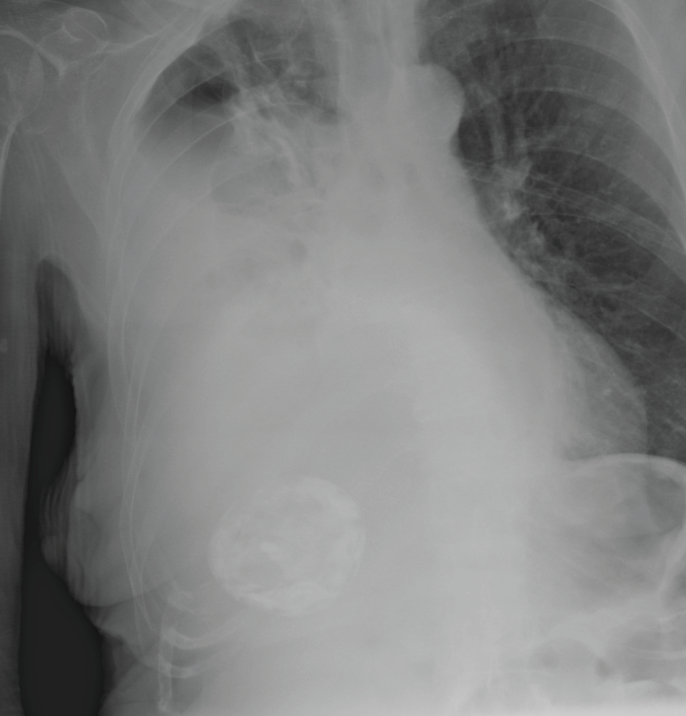
Figure 2: Hydropneumothorax clearly seen after removal of the nasogastric tube.
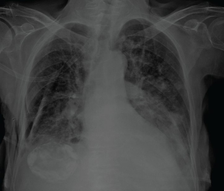
Figure 3: Resolution of hydropneumothorax after thoracentesis.
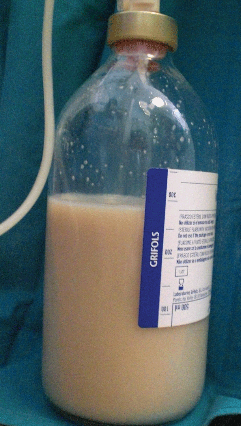
Figure 4: Enteral nutrition obtained after thoracentesis.