Twenty two years old female with left mammary gland lesion due to hyperprolactinaemia (32 ng/ml), nipple secretion and pain under pressure was referred to our Department. In the physical exploration, an approximate 2 cm solid nodule with regular edges and hypoechoic zones inside (suggestive of BIRADS3) was defined.
A metabolic evaluation of the mammary gland using Molecular Imaging (dedicated breast PET) after injection of 111MBq of 18F-FDG was performed.
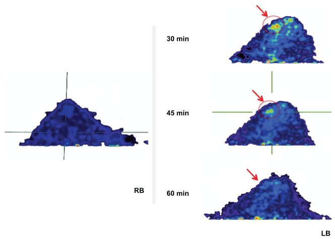
An early image after 30 minutes shows an area of homogeneous FDG uptake (hypermetabolic focus) placed within the periareolar area in IOQ with 4.54 SUVmax value. Radiotracer distribution in the rest of the parenchyma and in contralateral breast was within normal values. After 45 minutes, a considerable decrease of the uptake as well as significant decrease of the SUVmax (3.48) was found. Finally, after 60 minutes any foci of metabolic activity were detected and SUVmax values (2.4) are similar to those in the contralateral mammary gland and normal parenchyma. The authors confirm that Institutional Review Board approval (IRB) was obtained before the experiment (project no. 16462/RX663086, 24-July-2014).
In case of a malignant lesion, a PET study reflects an increase of FDG metabolism due to a high rate of tumor mass growth and proliferation versus normal tissues [1]. In tumoral cells, an increase of membrane transporters occurs which leads to internalization and accumulation of FDG into the cell [2,3]. Nevertheless, nothing is known about tracer wash-out in non-malignant lesion.
In this dynamic study a selected sequence of time (30, 45 and 60 minutes) was defined to evaluated the kinetic contrast and obtain the uptake curve for the analysis. Early image (30 min) reveals an increase of FDG uptake followed by a later image, which shows a loss in the metabolically activity (SUVmax from 4.54 to 2.7) close to the basal SUVmax in the healthy mammary gland. These results could be compatible with a non-malignant lesion and which a plateau curve type in relation to the kinetic contrast model.
This study gets us closer to a more precise diagnostic on mammary gland pathologies (especially in premalignant or non-malignant diseases [4]), highlighting the importance of detects false positives in early images. For the first time, a dedicated breast Molecular Imaging System (dbPET) [5] and dynamic study were associated to differentiate malignant and non-malignant pathology.

Figure 1: Transverse dbPET images at 30-45-60 minutes of galactocele on the left breast (LB) compared to healthy right breast (RB). Arrows show the area of greatest uptake, which disappears at 60 minutes.