IMAGE ARTICLE | VOLUME 2, ISSUE 1 | OPEN ACCESS DOI: 10.23937/2474-3682/1510024
Magnetic Resonance Images of the Structure and Tissue Organization of a Fetal Heart
Eleftheria Pervolaraki1 , Richard A Anderson2, Alan P Benson1 and ArunV Holden1
1School of Biomedical Sciences, University of Leeds, UK
2MRC Centre for Reproductive Health, University of Edinburgh, UK
*Corresponding author:
Eleftheria Pervolaraki, School of Biomedical Sciences, University of Leeds, Leeds, LS2 9JT, UK, E-mail: fbsepe@leeds.ac.uk
Published: January 25, 2016
Citation: Pervolaraki E, Anderson RA, Benson AP, Holden AV (2016) Magnetic Resonance Images of the Structure and Tissue Organization of a Fetal Heart. Clin Med Img Lib 2:024. doi.org/10.23937/2474-3682/1510024
Copyright: © 2016 Pervolaraki E, et al. This is an openaccess content distributed under the terms of the Creative Commons Attribution License, which permits unrestricted use, distribution, and reproduction in any medium, provided the original author and source are credited.
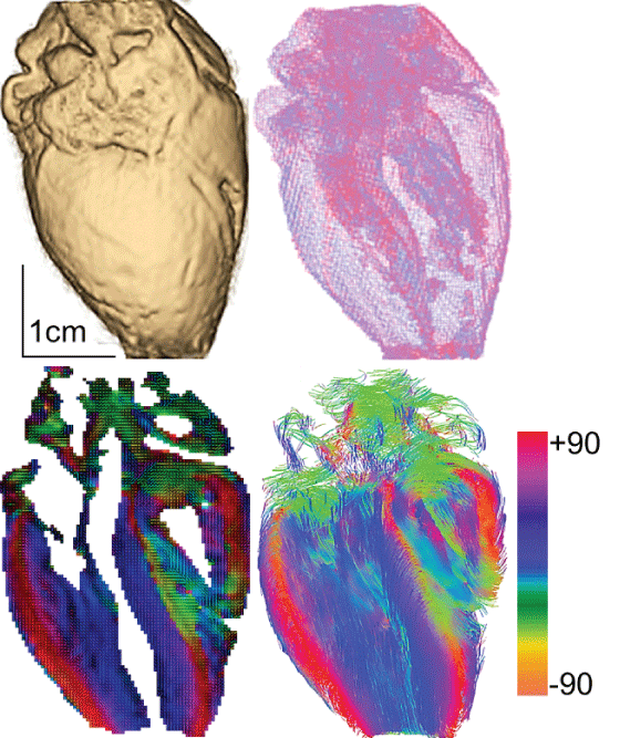
A 20 week gestational age human fetal heart was imaged at a 100 μm cubic voxel resolution, using a standard diffusion tensor magnetic resonance imaging (DTMRI) protocol in a 9.4 tesla Bruker NMR. The three-dimensional cardiac data set is visualized by its surface and a transparent wireframe (top panel). Bottom panel presents a long axis slice through the heart, where the local average myocyte orientation is visualized by a two-dimensional color map of the fibre helix angle, illustrating the smooth transmural change in fibre orientation, and by a cut through the 3D projection of the streamlines of the orientation vector field. Images were produced using open-source packages Para View (http://www.paraview.org/) and DSI Studio (http://dsi-studio.labsolver.org/).
