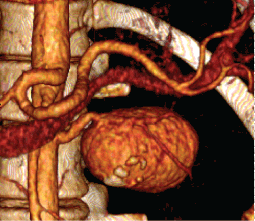
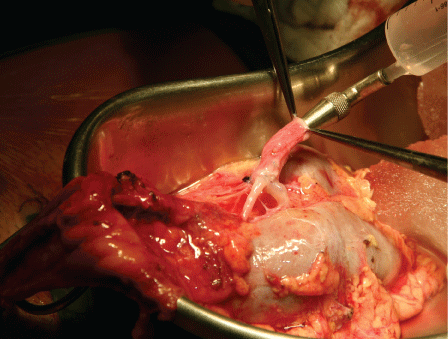
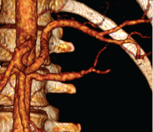
A 32-year-old woman presented with a one-year history of mild abdominal pain in the left upper quadrant and a palpable pulsatile abdominal mass on physical examination. The results of laboratory investigations, including serum urea and ceatinine levels, were unremarkable. Contrast enhanced Computed Tomography (CT) showed a large left renal artery aneurysm, measuring 5,0cm by 3,5cm, but no evidence of renal perfusion alterations or other vascular abnormalities (Figure 1). She had been previously submitted to an unsuccessful lendo vascular approach within tention to treat the aneurysm and preserve left renal perfusion. Because she was young and in good health, our purpose was to preserve left renal function and an open repair was adopted. The patient underwent a laparotomy with midline incision and the left kidney, left renal vein and artery were circumferentially mobilized from surrounding tissues. Left renal vein and artery were clamped and transected while the ureter was left intact and the ex-situ reconstructionwasperformedonthebodywall.Surfacecoolingandhypothermic renal perfusion with Euro-Collins solution (4 oC) was performed. Other authors have suggested that when more than 40 minutes of warm ischemia are required, the same assures to protect renal function should be instituted [1]. The aneurysm was then resected leaving the stump of the distal renal artery. The left hypogastric artery has been previously dissected and was used as autograft for renal artery reconstruction (Figure 2). Hypogastric autografts has been used in larger series and have important role in complex branched renovascular lesions [2]. Left renal vein was primarily and a stomosed and the kidney was returned to the original position. The patient was discharged in six days later, renal function remained unchanged and symptoms resolved. Duplex scan and contrast enhanced CT surveillance demonstrated left renal artery and vein patency (Figure 3).


