This is the case of a 17-year-old male who was saved from a penetrating thoracic gunshot wound by the extra-thoracic segment of a Nuss bar, placed 7 months earlier for treatment of a pectus excavatum defect. We discuss the significance of non-penetrating thoracic trauma and the workup involved.
The Nuss bar is a thin metallic prosthetic that is placed beneath a pectus excavatum defect to elevate the sternum. To date, there have been no reported cases of deflected bullets by such a device. There have similarly been no discussions of acute management of such injuries, potential sequellae, or the workup necessary to rule out such injuries.
A 17-year-old male with a history of pectus excavatum seven months status post Nuss bar placement presented to our trauma center with gunshot wound to the left chest wall. At the time of his injury, the patient was in a moving car that was hit by several bullets from an unidentified firearm. He was stable at the scene and transported to the nearest medical facility.
At the facility, the patient was complaining of left chest wall tenderness, but no other symptoms. Vitals were within normal limits and physical exam demonstrated a tall, thin patient, with a shallow penetrating wound to the left chest that was tender to palpation. Complete blood count, basic metabolic panel, and serum troponin were within normal limits. Electrocardiogram showed normal sinus rhythm with slight ST elevations in leads II, III, and aVF, with non-specific T wave inversion in V2. Two view chest radiographs demonstrated a single Nuss bar with bilateral stabilizing plates and no evidence of bullet fragments, rib fracture, pleural effusion, or pneumothorax (Figure 1). Chest CT with contrast showed induration of subcutaneous fat inferior and lateral to the left nipple, just anterior to the Nuss bar, with no evidence of fracture, vessel injury, or other intrathoracic injury (Figure 2). Due to the wound location and depth, it was noted by radiology that the Nuss bar likely reflected the bullet, preventing penetrating thoracic injury.
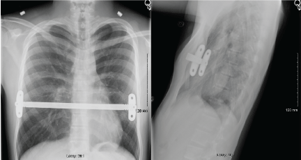 Figure 1: PA and Lateral radiographic imaging of the patient's Nuss bar status post gunshot wound with bilateral stabilizing plates visible. View Figure 1
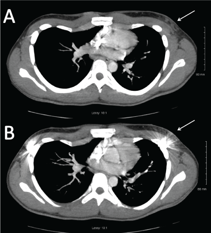 Figure 2: CT imaging demonstrating the bullet trajectory. Image A is approximately 6 mm superior to image B and demonstrates the soft tissue injury (white arrow) present in B that is scattered by metallic artifact from the Nuss Bar. These images provide a radiographic correlation for the clinical picture of a bullet wound superficial to the Nuss Bar. View Figure 2
Due to concern for cardiac contusion, he was transferred to our trauma center. Echocardiogram demonstrated a structurally normal heart without wall motion abnormalities. Repeat EKG was unchanged and the patient was discharged after overnight observation.
In our patient, there was clear evidence both by exam and on CT imaging (Figure 2) that the bullet struck the patient directly at the level of the extra-thoracic projection of the Nuss bar and was deflected. This is a fascinating case of bullet deflection by a metallic prosthesis but also a case that calls attention to the management and potential sequellae of non-penetrating gunshot wounds.
The Nuss bar is used for the surgical correction of pectus excavatum defects for either cosmetic or functional purposes [1]. The U-shaped metal rod is often a 1-2 cm, flattened appliance that is threaded under the concavity of the sternum using an introducer to avoid both the pericardium and internal thoracic vessels [1,2]. Either end of the Nuss bar exits the thorax and typically overlaps 2 ribs where it is sewn to the adjacent tissues for stability [1]. It is this extra-thoracic portion of the bar that was responsible for deflecting the projectile in our patient. To date, there are no reported cases in the literature of Nuss bar bullet deflections.
In our patient, management was straightforward and there were no occult injuries, but it is worth examining the potential sequellae. Non-penetrating injuries can cause internal damage, as evidenced in AA Razzaq's paper on the blunt injury to abdominal organs by surface gun shot wounds [3]. The mechanism, dubbed the "temporary cavity" effect is caused by tissue acceleration secondary to dispersal of a projectile's kinetic energy [3]. Shock waves generated through tissue were found to cause greater disruption of inelastic organs, such as the liver, when compared to flexible tissue like the lungs [3]. Domingo, et al. case of aortic dissection secondary to cavitary forces of a penetrating thoracic gunshot wound demonstrates the potential for this effect to occur within the thoracic cavity [4]. Although this effect was observed in penetrating trauma, Canon's article on Behind Armor Blunt Trauma catalogues the increase in blunt trauma secondary to bullet forces on light body armor [5]. In the case of our patient, the fragility of the Nuss bar as a form of body armor bore a significant risk of blunt trauma to the thoracic cavity with the potential for damage to heart, lungs, and significant vasculature.
Workup for a patient such as ours with a non-penetrating thoracic gunshot wound included chest radiographs to assess for gross fractures, pneumothoraces, or other traumatic injury, followed by CT with contrast for higher resolution imaging of structures including the heart, lungs, and vasculature. EKG and Troponin were performed to assess for blunt cardiac trauma. In the setting of our patient, the non-specific ST elevations and T wave inversion, otherwise normal in a healthy young individual, warranted further follow-up in the setting of traumatic injury and lack of prior study for comparison. Furthermore, pectus excavatum defects are often associated with syndromes such as Marfan syndrome or isolated findings such as aortic root dilatation [6]. In the setting of trauma, both Marfan syndrome and any congenital cardiac pathology may warrant further workup. According to a paper by Velmahos, et al. when taken together, TnI and EKG have sensitivity for blunt cardiac trauma of 100% but a positive predictive value of 34% [7]. Although the troponin was negative, the nonspecific EKG findings and concern for occult cardiovascular injury warranted overnight observation.
This case demonstrates a novel use of the Nuss bar as a form of light body armor. The bullet trajectory on CT and exam findings suggests that the bullet ricocheted off the extra-thoracic segment of the Nuss bar, potentially preventing penetrating injury to the thoracic cavity. In a patient such as this, the primary concern is for blunt trauma to the intrathoracic organs and warrants a workup that includes imaging of the organs and vasculature of the thorax along with EKG and serum troponin levels.