Trauma Cases and Reviews
Bilateral Osteochondrosis of Medial Cuneiform and Tarsal Scaphoid: A Case Report
Calderazzi F*, Carolla A and Ceccarelli F
Department of Surgical Sciences, Orthopaedic Clinic, University of Parma, Italy
*Corresponding author: Filippo Calderazzi, Department of Surgical Sciences, Orthopaedic Clinic, University of Parma, Via Gramsci 14 - 43100 Parma, Italy, E-mail: filippo.calderazzi@iCloud.com
Trauma Cases Rev, TCR-2-037, (Volume 2, Issue 2), Case Report; ISSN: 2469-5777
Received: March 30, 2016 | Accepted: May 24, 2016 | Published: May 27, 2016
Citation: Calderazzi F, Carolla A and Ceccarelli F (2016) Bilateral Osteochondrosis of Medial Cuneiform and Tarsal Scaphoid: A Case Report. Trauma Cases Rev 2:037. 10.23937/2469-5777/1510037
Copyright: © 2016 Calderazzi F, et al. This is an open-access article distributed under the terms of the Creative Commons Attribution License, which permits unrestricted use, distribution, and reproduction in any medium, provided the original author and source are credited.
Abstract
Osteochondrosis has been found in most of the bones of the body. We report a rare case report of bilateral osteochondrosis of medial cuneiform and tarsal scaphoid. A five year-old white child with a completely benign medical history presented with bilateral foot pain. Radiographical studies showed a bilateral irregular outline and an increased density of medial cuneiforms and tarsal scaphoids. Physical activity restriction and medical therapy appear to be sufficient for pain relief. This "syndrome" is self-limiting and it resolves in few months with relief of the foot pain.
Keywords
Osteochondrosis, Medial, Cuneiform, Tarsal, Scaphoid, Navicular
Introduction
Osteochondrosis has been found in most of the bones of the body. Harbin and Zollinger in 1930 first published a report on isolated osteochondrosis of the internal cuneiform [1]. Buchman in 1933 presented a study of 2 cases of osteochondrosis of internal cuneiform: in one of these there was a bilateral involvement of the first cuneiform and changes in the tarsal scaphoid, suggestive of osteochondrosis, such as slight condensation and some rarefaction in the center and an irregularity of the bony outline [2]. The same year Haboush presented the case of a male, four years old, with bilateral involvement of medial cuneiform and tarsal scaphoid [3]. Meilstrup in 1947 and O'Donoghue in 1948 presented similar cases [4,5]. To date, no other reports on this topic have been reported in literature. Considering this, we report the fifth case of bilateral osteochondrosis of medial cuneiform and tarsal scaphoid. Purpose of this short report is to invite the reader to consider also this rare pathology, because the correct diagnosis influences the treatment choice.
If this pathology is not correctly diagnosed and treated, as all other osteochondrosis, it can potentially lead to bone deformity, also if in literature has not been reported yet.
Case Report
A white five year-old male with a completely benign medical history, presented with bilateral foot pain, persistent after eight weeks from first onset. The pain arose the first time during overload playful sports activities. No history of previous trauma, recent infections, or puncture wounds were noted. There was no clear family history of osteochondrosis and there weren't any correlations with climatic changes or with episodes of freezing.
.
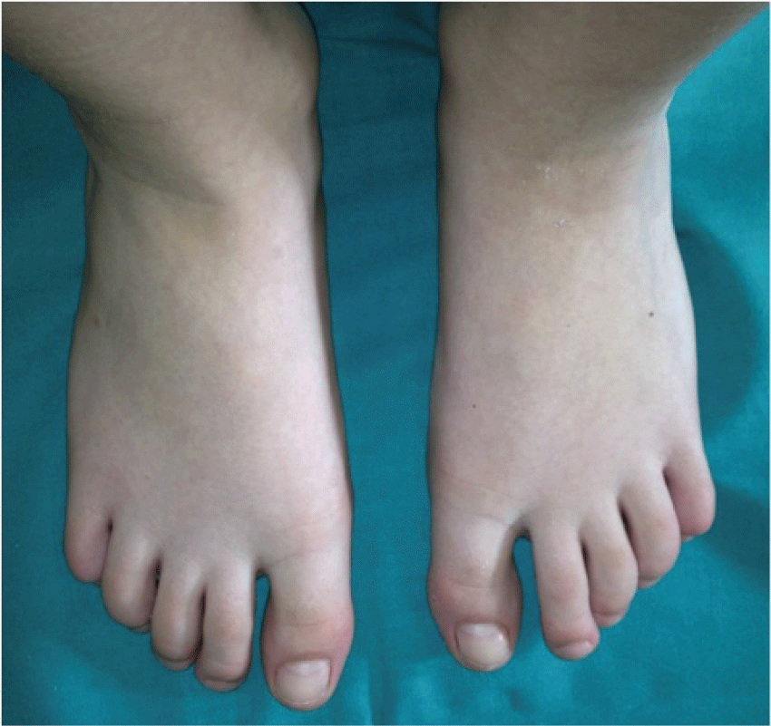
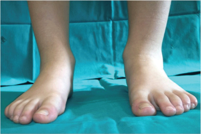
Figure 1: Clinical picture in dorsoplantar view at first examination - significant pathological morphologic changes are not clinically present.
View Figure 1
.
Figure 2: Clinical picture in frontal view at first examination - significant pathological morphologic changes are not clinically present.
View Figure 2
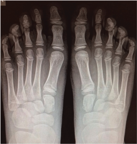
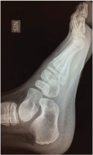
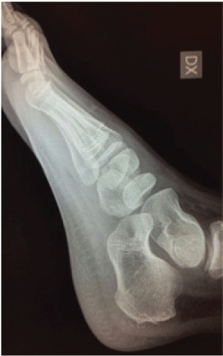
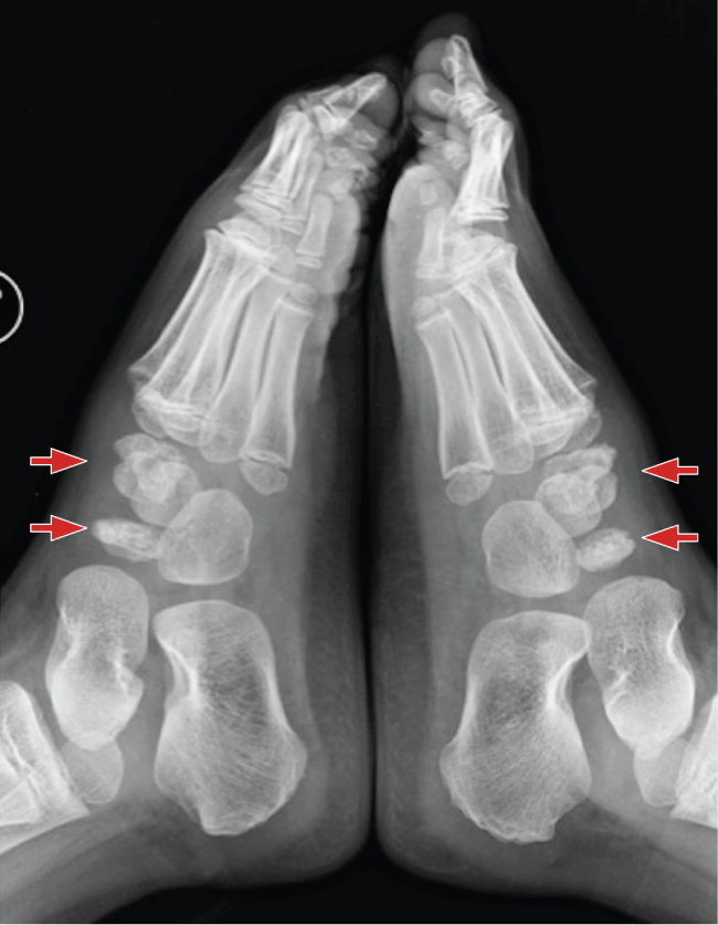
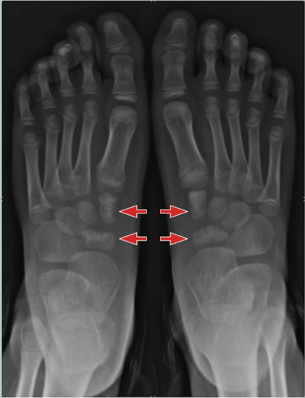
The patient complained of bilateral pain of mid medial foot after physical activities, mainly at the end of the day. The first onset of symptoms was described as a discomfort, worsened with activity and relieved with rest. Physical examination reflected an otherwise normal healthy young boy, with normal weight. No edema, soft-tissue swelling, or discoloration was observed at the feet (Figure 1 and Figure 2). Full range of motion of all joints was accomplished without any pain. Careful palpation of the medial dorsal aspect of the midfoot reproduced the patient's pain. Laboratory tests like a complete blood count, erythrocyte sedimentation rate (ESR) and C-reactive protein (CRP) were normal. Radiographical studies showed a bilateral irregular outline and an increased density of medial cuneiforms and tarsal scaphoids (Figure 3 and Figure 4). No significant changes were noted in the other bones of the feet. MRI was not carried out. The patient was treated with acetaminophen, a mild analgesic, and limitation of physical activity. He was able to carry out normal activities with the help of custom orthosis to support the arch in order to reduce the pain, but he had to refrain from sports.
.
Figure 3: X-ray lateral view at first examination (Higher arrows: osteochondrosis of the medial cuneiform; Lower arrows: osteochondrosis of tarsal scaphoid).
View Figure 3
.
Figure 4: X-ray dorsoplantar view at first examination (Higher arrows: osteochondrosis of the medial cuneiform; Lowest arrows: osteochondrosis of tarsal scaphoid).
View Figure 4
We put the patient through monthly follow-up. He had gradual resolution of the symptoms and was symptom-free at ten months follow-up. Radiographic evaluation performed at this time showed complete reconstitution of the size and trabecular patterns of the medial cuneiforms and tarsal scaphoids (Figure 5, Figure 6A and Figure 6B). The clinical picture had remained unchanged one year later.
Discussion
Osteochondrosis have been defined as idiopathic "syndromes", characterized by a disorder of enchondral ossification that occurs in the area of a previously normal growth mechanism [6]. There are more than 75 bones or portions of bones that have been documented as sites of occurrence of this growth disturbance [7]. Although osteochondrosis of the tarsal and metatarsal bones are common in children, involvement of the cuneiform bone is rare [8].
A comparison of this case with the previous ones encompasses a marked similarity in the findings: no history of trauma, masculine sex, age between 4 to 6, normal laboratory test, pain (described as "discomfort") worsening with activity and relieving with rest [1-5]. It is not possible to compare the activity level of our patient with what has been described in previous reports. Literature is not clear about the role of microtrauma as an underlying causative factor of osteochondrosis [1-5]. However some Authors suggest, being the osteochondrosis a vascular disorder, that microtraumas can theoretically increase the process of bony disarrangement [9].
In relation to literature that we have available, the age of onset more likely varies from 4 to 6 years and may affect the diagnosis [8].
Based on our experience and in accordance with the literature findings, bilateral osteochondrosis of internal cuneiform and tarsal scaphoid appears to be a self-limited disorder, that may improve in a relatively short time and without pathological sequelae.
Radiographic evaluation, if performed in the early stages, may demonstrate patchy increase in density, nodularity and fragmentation of the involved bone [10]; a dorsoplantar view gives enough details to a clinician, without need of other special projections.
Over a period of 6 to 24 months, the bone usually regains its normal size, density, and trabecular markings [7,8]. The differential diagnosis is rather limited. Other diseases that should be considered, at least from a radiographic standpoint, include the Ewing's sarcoma, osteomyelitis and eosinophilic granuloma, frostbite [11]. Careful history-taking and accurate physical examination usually exclude all other diagnoses [6-8]. MRI is usually not necessary, as it does not add further significative informations to clinical and radiographic picture [8]. Moreover, given the young age of the patient, MRI should be performed under sedation. Some recent studies have pointed out that high-field MRI is able to detect early changes of osteochondrosis in animal models; however nowadays this method is not still applicable in daily practice [12].
Conversely, failure to recognize this "syndrome" may lead to treat this lesion with an aggressive approach (open biopsy or excision) that may be unnecessary and potentially harmful to the patient [7].
The therapy should consist on non-surgical, symptomatic-related treatment. Acetaminophen or any other analgesic well tolerated by the children is usually the only drug required and is adequate to relieve pain [6,7].
Physical activity restriction and medical therapy appear to be sufficient for pain control. A biomechanical support by means of foot orthoses, padding and strapping can also be useful [6]. Immobilization in a plaster cast for a short period is infrequently used [7]. Since this is a self-limiting disease, we do not agree with treatments that include a prolonged immobilization in a plaster cast.
Conclusion
This "syndrome" is self-limiting and it resolves in few months with relief of the foot pain. Physical activity restriction and medical therapy appear to be sufficient for pain relief. Indeed, an important aspect of the management of this "syndrome" is to reassure the parents that the condition is likely to be self-limiting and to resolve with time.
Level of Clinical Evidence
The level of clinical evidence is 4. Authors declare that they have no conflict of interest. The study was authorized by the ethical committee of the authors' institution.
References
-
Harbin M, Zollinger R (1930) Osteochondritis of the growth centers. Surg. Gynec. Obstet 51:145-161.
-
Buchman J (1933) Osteochondritis of the internal cuneiform. J Bone and Joint Surg 15: 225-232.
-
Haboush FJ (1933) Bilateral disease of internal cuneiform bone with associated disease of right scaphoid bone (Köhler's). JAMA 100: 41-42.
-
Meilstrup DB (1947) Osteochondritis of the internal cuneiform, bilateral; case report. Am J Roentgenol Radium Ther 58: 329.
-
O'Donoghue AF, Donohue ES, Zimmerman WW (1948) Bilateral osteochondritis of the tarsal navicular and the first cuneiform; a case report. J Bone Joint Surg Am 30A: 780.
-
Kose O, Demiralp B, Oto M, Sehirlioglu A (2009) An unusual cause of foot pain in a child: osteochondrosis of the intermediate cuneiform. J Foot Ankle Surg 48: 474-476.
-
Leeson MC, Weiner DS (1985) Osteochondrosis of the tarsal cuneiforms. Clin Orthop Relat Res 260-264.
-
Atbasi Z, Ege T, Kose O, Egerci OF, Demiralp B (2013) Osteochondrosis of the medial cuneiform bone in a child: a case report and review of 18 published cases. Foot Ankle Spec 6: 154-158.
-
DiGiovanni CW, Patel A, Calfee R, Nickisch F (2007) Osteonecrosis in the foot. J Am Acad Orthop Surg 15: 208-217.
-
Resnick D (1981) The osteochondrosis. In: Resnick D, Niwayama G, Diagnosis of Bone and Joint Disorders. WB Saunders, Philadelphia, 2874.
-
Pettit MT, Finger DR (1998) Frostbite arthropathy. J Clin Rheumatol 4: 316-318.
-
Tóth F, Nissi MJ, Wang L, Ellermann JM, Carlson CS (2015) Surgical induction, histological evaluation, and MRI identification of cartilage necrosis in the distal femur in goats to model early lesions of osteochondrosis. Osteoarthritis Cartilage 23: 300-307.


