Acute mesenteric ischemia is a rare complication of thromboembolic diseases in the mesenteric vessels, which was noted to occur due to the hypercoagulable circulation associated with pregnancy. Additionally, the early detection and treatment of such disease prevents further deterioration of the mother's condition, thus ensuring a favorable outcome for both the mother and the fetus. This paper presents a case report of acute mesenteric ischemia in a 9 weeks pregnant female, which was diagnosed late due to the non-specific presentation of the disease, yet was adequately treated with an acceptable result.
Acute mesenteric ischemia, Mesenteric venous thrombosis, Intestinal resection
Mesenteric venous thrombosis (MVT) is a rare cause of intestinal ischemia, which when happens in pregnancy is life threatening to both the mother and the fetus making early diagnosis crucial. However, due to the vague abdominal symptoms during pregnancy and the preferability to avoid radiological investigations, diagnosis of MVT becomes challenging [1].
A 31-year-old female, gravida 4 para 3, 9 weeks gestational age, presented to the ER complaining of severe lower abdominal pain. The patient was admitted due to the onset of vomiting which was diagnosed as hyperemesis gravidarum.
On examination, the patient showed severe pelvi-abdominal tenderness. Ultrasound was performed showing single viable fetus with average crown-rump length (CRL) ± 9 weeks, and moderate pelvi-abdominal collection noted up to Morison pouch. BP was 90/60 and the pulse was 140/min and faint.
Complete blood picture was done showing: haemoglobin level of 11.3 gm/dl, Platelet count of 226,000/µl, and White blood cell count of 16,000/µl.
U/S guided aspiration from the Morison pouch was performed reviling bloody fluid.
The decision to perform a midline exploratory laparotomy was taken. Vital signs before receiving general anesthesia showed: BP: 90/60 and pulse: 130/min. After opening the skin, subcutaneous tissue, muscle and peritoneal layers, gangrenous bowel loops were noted and 750 cc of bloody fluid collection was suctioned. At first application of hot foments to the gangrenous loops was applied several times, however no signs of revitalization were noted. A segment of 2.5 meters of small bowel loops, 40 cm apart from the duodenojejunal junction, was resected and end-to-end anastomosis was performed. The patient was then transferred to the ICU for postoperative observation.
Intraoperative photographs were taken demonstrating the gangrenous non-viable small bowel loops before resection, shown by the clear demarcation in color change (Figure 1). During resection and anastomosis, a mesenteric venous branch was found to have a localized firm segment. Upon dissection of this vessel segment, a 1.5 cm venous thrombus was discovered occupying the lumen and was extracted (Figure 2).
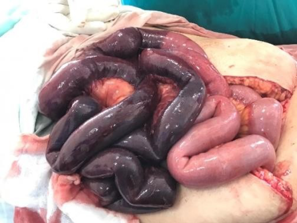 Figure 1: Loops of gangrenous bowel before resection.
View Figure 1
Figure 1: Loops of gangrenous bowel before resection.
View Figure 1
 Figure 2: Thrombus extraction from a mesenteric vessel.
View Figure 2
Figure 2: Thrombus extraction from a mesenteric vessel.
View Figure 2
Three days later the patient was stable, no postoperative complications presented, and she was discharged from the ICU and advised to return for constant follow ups. The course of the pregnancy continued without any complications. At 34 weeks, the patient presented with preterm labor pain (PTLP) and upon consultation a decision was made to terminate the pregnancy. A cesarean section operation was performed, the baby was delivered and transferred to the NICU and was discharged four days later. Both the mother and the baby were then stable and were discharged from the hospital.
Mesenteric Venous Thrombosis was first differentiated and described as a cause of acute mesenteric ischemia by Warren and Eberhard in 1935 [2]. A lack of recent epidemiological studies was noticed, however in 2015 a retrospective study reported an incidence rate of acute mesenteric ischemia of 7.3/100,000 person per year. Acute mesenteric ischemia occurs due to arterial occlusion in 65% of cases, venous occlusion in 28% while 7% were due to non- occlusive causes [3].
The hypercoagulable state associated with pregnancy puts the female at an increased risk of developing thromboembolic diseases [4]. In fact, it was documented that the hypercoagulable state in normal pregnancy causes an approximate increase in incidences of venous thromboembolism (VTE) by 6-folds [5].
In 2017 a few cases of acute mesenteric ischemia in pregnancy were published, some cases showed some sort of pathology that could have been a predisposing factor to the thromboembolic disease as antiphospholipid syndrome [6] or following surgery [7]. However other cases showed no additional predisposing factor rather than the pregnancy itself [8,9].
With a rate of 1 in every 635 pregnancies, acute abdomen during pregnancy was found to be caused by both obstetrical, non-obstetrical, gynecological and non-gynecological causes making its management challenging [10,11]. Therefore, relying solely on the clinical presentation and abdominal examination for the diagnosis of non-obstetrical and non-gynecological causing of an acute abdomen in pregnancy is to some extend inconclusive due to the physiologic changes associated with pregnancy that can mimic some types of pathology [11]. And when it comes to the use of radiology (e.g. CT) to aid in reaching the diagnosis, the Society of Obstetricians and Gynecologists recommend the use of CT and gadolinium contrast agents, when the benefits outweigh the potential risks, through evidence released in 2014, level III-C [12].
Early diagnosis and treatment of acute intestinal necrosis caused by MVT is critical for the survival of both the mother and the fetus. However not only does the various causes of acute abdomen in pregnancy, make the early diagnosis of AMI due to MVT remain challenging, but also the reduced ability to use computed tomography (CT) which remains a golden standard in the diagnosis of MVT [13,14]. Nevertheless, late presenting cases could also be rescued in the hands of well-trained medical personal. Owing to the rarity of such diagnosis, gastrointestinal lesions should be more often investigated in gravid females with acute abdomen or vague gastrointestinal symptoms.