Penetrating injuries usually affects non-dressed parts of body. Most of them treated by patient itself without any intervention. But some of them must be examined and treated by a specialist such as major injuries, minor injuries affecting the important structures and curved foreign bodies. Curved injuries can be made by a curved wire or fishhooks. Fishhooks take an important part of this injuries because of mechanism used in distal part of fishhooks. In this article we discuss two injuries resulted from curved materials and its management. One patient was injured with a fishhook and the other patient was injured with curved wire. We discuss patients and also review the literatüre related with such injuries.
Fish hooks, Foreign body, Curved
Penetrating trauma to the head and neck is relatively rare, representing around 1-2% of all traumas but when encountered can have devastating clinical consequences. Penetrating oral cavity injuries are mostly seen in young children between 2-6 years-old. This period have risks because of they tend to put or carry objects in their mouths for oral stimulation, and also they can easily loose their balance.
Injury of important structures in the head and neck region and injuries in the airway may result in serious morbidities and even mortality. Because of these complications prompt and careful intervention is necessary.
A 8-years-old male was injured with fishhook from his tounge during fishing. Fishhook embedded in his tounge during he was throwing fishing line. There was slight introral bleeding but there were no any respiratory distress and neurologic findings. The patient was evaluated by a practioner in emergency room and referred to the otorhinolaryngology service. He had good medical history without admission to the hospital for any reason. His tetanus vaccination was checked and found to be effective.
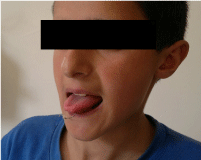
Patient was sitting without any signs of bleeding or respiratory distress. The fishhook was in the middle part of anterior inferior tounge. The tounge was a little edematous because of prior interventions for removing fishhook. There was a little amount of blood in the floor of mouth (Figure 1).
 Figure 1: Fishhook is seen embedded in the inferior part of tounge.
View Figure 1
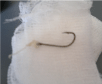
The patient was taken to the operating room for possible complications. The fishhook was embedded in its last curve about 4 milimeters. The fishhook was removed without any complication (Figure 2). At the time of control after 1 week, the laceration had healed well.
 Figure 2: Removed fishhook.
View Figure 2
A 60-year-old-man who presented to Emergency Departement 2 h after accidentally embedding a curved wire in his right outer ear canal. It had become more embedded when patient and doctors in emergency departement were attempting to remove it. Before presenting a doctor in emergency service tried to remove but they could only increase edema in outer ear canal instead of removing it. There was not any bleeding at time of presentation. The patient complained of mild hearing loss and pain in ear but he did not noticed any other additional symptoms. His tetanus immunization was done in emergency departement.
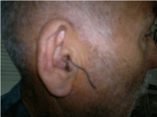
On physical examination, a wire was getting out approximetely 8 centimeter and he did not know how long it was (Figure 3). Right outer ear canal was edematous and nearly obstructed with swelling. There was a minimal haemorrhagic discharge from affected ear. He had no any history of ear disease before. For extension we planned a CT scanning for possible ear drum or middle ear injury.
 Figure 3: Curved wire embedden in right outer ear canal. Canal edema and discharge is also seen in figure.
View Figure 3
There was not any middle ear extention of wire and it was seen that wire was curved at the end. It could not be extracted easily because of curved structure.
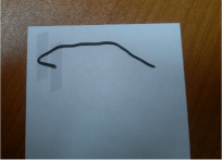
After proper cleaning and dressing, local ansethetic infiltration was made. With microscopic evaluation wire was removed without any complication (Figure 4). There was outer ear canal laceration in proximal outer ear canal and edema in outer part of ear canal. Topical ear drops were prescribed with oral antibiotic pills. After 1 week there was not any signs and symptoms of edema and otitis externa as well as any hearing disorder.
 Figure 4: Removed foreign body from right ear canal.
View Figure 4
Management of foreign bodies needs attention. Especially fishhooks needs more attention because of barb in the curved distal part that resistant for simple removal. Direct forces for removing fishhooks may result in tissue damage and can cause complications like bleeding or pain. Different fishhook injuries in head and neck region such as eyeball, hypopharynx, skull, tounge and soft palate have been reported [1-5].
Those which has proximity to vital structures like vessels, nerves or injury to the special organs like orbita, oropharynx need more attention and must be managed by a specialist [6]. Complications of this kind of important structures may compromise in irreversible results such as vision lost.
During taking history; injury time and the type of injury, attemps for removal must be obtained as well as tetanus immunization history. Related symptoms must be checked during examination.
İmportant structures around injury field must be checked. Also the distal end of fishhook must be evaluated for possible bone or cartilage involvement. Most fishhook injuries are in hand, face, head or upper extremity [7].
If fishhook was not detached from fishingline or other instruments, it must be seperated from distal fishing line for preventing possible complications. Generally in head and neck region fishhook are embedded to the skin or mukozal tissue but in extremity X-ray scanning may be helpfull.
Surgical interventions always starts with proper cleaning and dressing. Surgical removal of foreign bodies starts with local care. If fishing line is not detached from fishhook, fishing line must be seperated from fishhook as possible as near to the fishhook for preventing possible complications and more ease manipulation. Local anesthetic infiltration may be necessary in some cases especially in patients with low pain threshold or areas that needs more attention.
There have been several techniques described for removing curved foreign bodies with barbs. All of them designed for preventing further injury of barb during removing. There are few articles describing methods and its success rates [8]. Type of fishhook, location of injury and the depth of fishhook in tissue affects the technique selection. Sometimes combinations may be necessary.
In string-yank method, a string is placed on the curve of fishhook and makes a parallel power tension to the long part of hook. While pulling through a string a downward pressure is applied to the long part of hook so barb displaces into the hooks curved embedded part and moving it back through way of cutting. With using this technique 17.5% fishhooks can be removed [9]. We also used this technique in our case for removal.
Retrograde method is the least effective technique while it is the most easiest technique. This method is like string-yank method but this method does not use string. Instead of string manual pressure is applied to the hook first downward and after both down and back to the long axis of hook. It has same principals with string-yak method but it is more selected in more superficially located hooks. This method is also found effective in 17.5% of patients.
Needle cover method has a principal that covering the barb of the hook within the hollow point of a needle. 18-gauge needle is generally preferred for this method. A needle tip is moved toward to the barb of fishhook for covering it. When barb is covered with a tip of needle it is aimed to decrease the pain and tissue damage effect of barb. Once this covering can be maintained, the hook and needdle removed together out. This method reported to have a 7% effectiveness rate.
Advance and cut method includes advancing and cutting the barb. Fishhook is advanced and when the barb is get out of tissue barb can be clipped off. When the barb clipped off it can be removed easily in retrograd pathway. If there are multiple barbs this method can be usefull. It is reported to be effective in 58% of cases [9]. Its limitaion is hard tissues where the hook cannot be advanced to be cut, such as finger where bone or fingernail is involved.
Advance without cutting is usefull if hook can be extracted from a second exit. This method used in very limited case reports especially in eyelid [10].
Cut it out method means an incision is made at the entry point of the hook and hook is taken out. This method has more morbidity for scarring and tissue damage and preferred as a rescue technique if other methods fail. Cut methods are generally not preferred in head and neck region for esthetic results.
Most of oral injuries are minor and can be managed with follow-up or minor interventions. However it must be kept in mind that this kind of injuries can have lethal acute or delayed complications especially which involves and affects upper respiratory tract [11]. As a result of close proximity of oral cavity to the important structures like parapharyngeal and retropharyngeal space, many serious complications can result from injuries of oral cavity.
Usual foreign bodies in ear are cotton fragments 24.06%, Insects 22.5% and other soft structures [12]. Curved foreign materials are very rare.
Therapeutic success depends on a number of factors, but there is no strong evidence to indicate one specific removal method over others.
Generally wound is left open. We recommend oral antibiotic treatment and antibiotic ointments or drops although there is not a consensus in this topic.
Management of curved foreign bodies are challenging procedures in otorhinolaryngology. Patients should be managed with special attention to airway protection, nursed in the upright position, and all potentially distressing interventions should be minimized. Prompt removal of the curved foreign body should be performed under direct vision to prevent further soft tissue damage. After removal of curved foreign bodies, the wound must be checked for possible retained foreign bodies in wound. Tetanus immunization must always be checked in all foreign body cases.
None.
None.
We clearly declare that all the listed authors have participated sufficiently in the preparation of the manuscript and take responsibility for the content of the manuscript. Each author has contributed to study design, harvest and interpretation of the data, drafting and revising it for intellectual content.