Journal of Otolaryngology and Rhinology
Inadvertent Vocal Fold Polypectomy During Tracheal Intubation; A Case Report
Sean Keane1*, Patrick Dillon1 and John E Fenton2
1Department of Anaesthesia, University Hospital Limerick, Ireland
2Department of Otorhinolaryngology, University Hospital Limerick, Ireland
*Corresponding author:
Dr. Sean Keane, Specialist Registrar in Anaesthesia, University Hospital Limerick, Co. Limerick, Ireland, Tel: 0035-3877-732659, E-mail: sean.e.keane@gmail.com
J Otolaryngol Rhinol, JOR-3-028, (Volume 3, Issue 1), Case Report
Received: June 23, 2016 | Accepted: January 18, 2017 | Published: January 21, 2017
Citation: Keane S, Dillon P, Fenton JE (2017) Inadvertent Vocal Fold Polypectomy During Tracheal Intubation; A Case Report. J Otolaryngol Rhinol 3:028.
Copyright: © 2017 Keane S, et al. This is an open-access article distributed under the terms of the Creative Commons Attribution License, which permits unrestricted use, distribution, and reproduction in any medium, provided the original author and source are credited.
Abstract
A 48-year-old male presented for microlaryngoscopy and vocal fold polypectomy, which was complicated by polyp displacement into the lower respiratory airway during direct laryngoscopy and tracheal intubation. It is the first reported incidence of such a case. Following preoperative assessment and preparation, anaesthesia was induced and the airway secured using direct laryngoscopy and tracheal intubation with a microlaryngoscopy tube (MLT), which was reported uneventful. On microlaryngoscopy, the surgeon who had diagnosed the polyp in the outpatient clinic noted the polyp had disappeared. It was determined that the polyp had been sheared by the MLT. A fibreoptic scope was passed via the MLT, showing the polyp at the entrance to the right main bronchus. The polyp was successfully extracted via rigid bronchoscopy. The subsequent surgery and post-operative recovery were otherwise uneventful. Complications of bronchial occlusion were avoided by early recognition of the issue. Case reflection identifies many potential learning points. Head, neck and airway lesions add a dimension of complexity to anaesthetic practice. Preoperative assessment requires review of relevant endoscopic and radiographic images, and case discussion with the surgeon to identify potential issues. The location, size and vulnerability of any potentially complicating lesions must be known. The vocal folds and documented lesions should be visualised throughout intubation, and excess force during intubation avoided. This may require the use of alternative devices or techniques. Preoperative laryngoscopy by the operating surgeon or photo-documentation of the lesion may be necessary for microlaryngoscopies where the original physician who noted the abnormality cannot be present in the operating theatre.
Keywords
Microlaryngoscopy, Direct laryngoscopy, Polypectomy, Iintubation, Aspiration, Complication, Tissue trauma, Vocal fold
Summary
A 48-year-old male presented for microlaryngoscopy and vocal fold polypectomy as a day-case procedure. Anaesthesia was induced and the airway secured via direct laryngoscopy and tracheal intubation using a microlaryngoscopy tube (MLT), which was reported uneventful. On microlaryngoscopy the polyp had disappeared, leaving a small residual area of bleeding. It was determined that the polyp had been sheared by the MLT at intubation. A fibreoptic scope was passed via the MLT, showing the polyp at the entrance to the right main bronchus. The MLT was removed and rigid bronchoscopy performed with successful removal of the polyp. Surgery and post-operative recovery were otherwise uneventful. Complications of bronchial occlusion were avoided by early recognition of the issue. Clinic notes and relevant imaging should be reviewed pre-operatively, and potential airway issues discussed with the surgeon. Additional attempts should be made to visualise the vocal folds throughout tracheal intubation. Awareness of this potential problem may prevent similar events occurring in the future.
Introduction
Vocal fold polyps are common, typically unilateral and have a broad spectrum of appearances. They can present as haemorrhagic to oedematous, pedunculated to sessile, and gelatinous to hyalinise. The polyps are benign in the vast majority of cases, and tend to arise from phonotrauma or an area of previous haemorrhage [1]. Alteration in voice is the most common presentation, with symptoms including hoarseness, low pitch and reduced phonation. Therapy can be operative or non-operative, with the operative approach being deemed appropriate in this case due to clinical presentation, polyp morphology, and potential for malignant change.
Microlaryngoscopy is a common procedure that every anaesthetist will be familiar with. As with all forms of airway surgery, it is a procedure that requires a great deal of cooperation between the anaesthetist and surgeon. The anaesthetist is required to provide an immobile and unobstructed operative field, in conjunction with the usual requirements of providing oxygenation, carbon dioxide removal, adequate anaesthesia, and swift return of consciousness and airway reflexes postoperatively. Many techniques exist to provide anaesthesia for this procedure, including direct laryngoscopy with tracheal intubation, low-frequency jet ventilation via suspension laryngoscope, and high-frequency jet ventilation via a subglottic cannula.
Complications associated with this procedure are rare, and the incidence of issues related to direct laryngoscopy and tracheal intubation are scant in the literature. This report describes a case of tracheal intubation causing unrecognised shearing of a relatively small pedunculated vocal cord polyp. The inadvertent polypectomy was recognised by the surgeon, and it was located at the entrance to the right main bronchus. The polyp was successfully removed without incident, but many potential learning points were identified upon case reflection.
Case Report
A 48-year-old male presented to the ENT service with persistent hoarseness. He was an ex-smoker with a 40 pack year history, and past medical history was otherwise unremarkable. A flexible nasendoscopy performed 4 weeks previously by the consultant ENT surgeon at the outpatients clinic revealed a relatively small bilobed, pedunculated polyp on the anterior third of the left vocal fold close to the anterior commissure. A CT-neck and thorax were ordered, and the patient booked for urgent microlaryngoscopy with excision of vocal fold polyp as a day-case procedure.
The anaesthetic preoperative visit took place on the morning of surgery and did not highlight any concerns. CT-neck was reviewed by the operating surgeon and revealed no abnormality of note. Surgical outpatient notes were not reviewed prior to induction of anaesthesia, and the vocal fold pathology likely to be encountered was not discussed with the ENT consultant present. Anaesthesia was induced using fentanyl, propofol and rocuronium. Direct laryngoscopy was performed by a senior registrar using a macintosh 4 blade. The trachea was intubated using a microlaryngoscopy tube (MLT) size 6.0, with no reported difficulty. Mechanical ventilation and oxygenation were straight forward at this time.
Microlaryngoscopy was performed by the consultant surgeon who performed the diagnostic flexible nasendoscopy in the outpatient clinic. Visualisation of the larynx revealed a small area of bleeding on the left anterior vocal fold, with no sign of the bilobed pedunculated polyp. The appearance was immediately recognised as being different to what was seen in the outpatient setting. A detailed examination of the larynx and pharynx could not locate the polyp, and it was surmised that it was sheared off by the MLT at the time of intubation, and likely entered the lower respiratory tract.
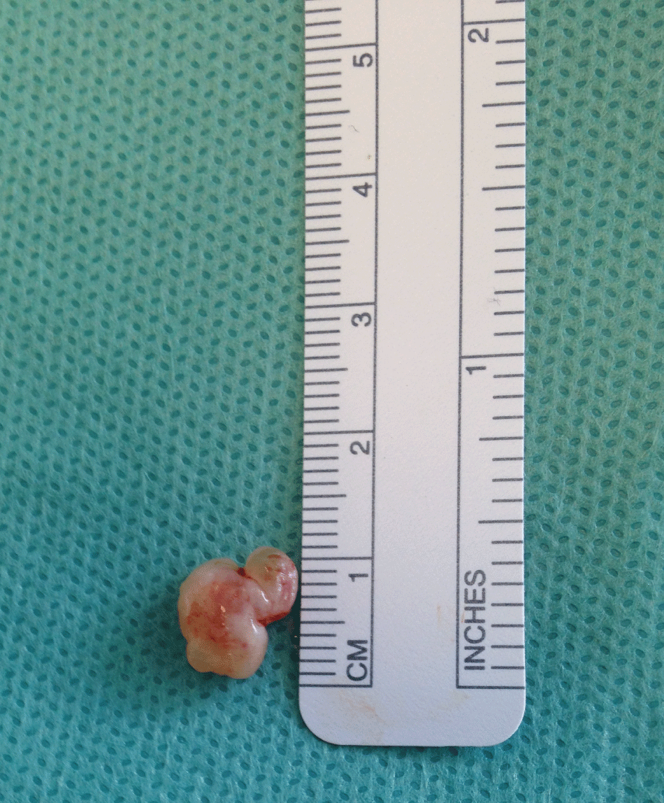
A detailed discussion was held between the consultant surgeon and anaesthetist to form an appropriate management plan. A fibreoptic scope was passed via the MLT, with the polyp being identified at the entrance to the right main bronchus. Deliberation concluded that an attempt to remove the polyp using suction via the fibreoptic scope may exacerbate the problem by dislodging the polyp further down the right main bronchus. The MLT was subsequently removed and rigid bronchoscopy performed. The polyp was successfully extracted via the rigid bronchoscope using a forceps, and the bronchi appeared patent. The MLT was re-inserted and surgery completed with no further issues. Gross examination of the polyp revealed a relatively small, bilobed polyp approximately 10 millimetres in diameter with a benign appearance (Figure 1). The polyp was sent to the laboratory for histology. There were no clinical signs or symptoms of lobar collapse, and no further investigations were arranged. The post-operative recovery period was uneventful.
The intra-operative events were discussed with the patient and concerns of further complications as a result of the incident were allayed. He was discharged home on the day of surgery as originally planned. At routine surgical outpatient follow-up four weeks later the patient was very well and histology was consistent with a benign nodule.
Discussion
This is the first reported case of a vocal fold polyp being sheared during insertion of an endotracheal tube. The sequences of events in this case strongly indicate direct laryngoscopy and tracheal intubation displacing the polyp into the conducting airway. The presence of bleeding on the left anterior vocal fold at the time of initial microlaryngoscopy support this, and further imply tissue trauma as a direct result of anaesthetic intervention. Microlaryngoscopy with vocal fold polypectomy is a common surgical procedure, and it is remarkable that this is as far as we can ascertain, the first incidence of such a complication. The pedunculated polyp in this case was likely more susceptible than other forms of polyps to such an injury, but it stands to reason that anaesthetists give greater consideration to the occurrence of such an issue in routine practice.
Adverse issues associated with direct laryngoscopy are uncommon, with cases of airway compromise exceedingly rare [2]. An extended search of the literature failed to find any cases of foreign body in the lower respiratory tract as a result of direct laryngoscopy and tracheal intubation, though there is a case of layrngoscope bulb ingestion in the paediatric setting [3]. A broader search to include the issues associated with microlaryngoscopy reveal mucosal damage [4], dental trauma and lingual nerve injury [5], as being potential complications, but no reports of tissue aspiration. Additional peri-operative causes of iatrogenic foreign body in the airways are rare in the literature. Surgical instrument failure leading to inadvertent loss of bronchoscopy instruments [6], and an aspiration of a fractured portion of microlaryngoscopy equipment [7], into the tracheobronchial tree, has been reported. A case of inadvertent injection of a nozzle tip through the vocal cords into the right main bronchus during attempted administration of topical anaesthesia to the cords using 10% lidocaine via a long spray delivery tube has also been described [8], recognised equipment failure is a common theme in many cases of peri-operative foreign body aspiration, and thus represents a vastly different mechanism of injury to traumatic tissue displacement described in this case.
The polyp in this case measured approximately 10 millimetres in diameter. Bronchial obstruction would have likely occurred at a point between the right main bronchus and right lower lobe bronchus, and could have dramatically impacted events in the intra-operative and postoperative setting. Intra-operative issues may include hypoxemia due to shunting, and high airway pressures, while issues in the postoperative setting can encompass atelectasis, lobar collapse, repeat surgical intervention, infection and prolonged mechanical ventilation. The severity of such consequences often hinges on promptness of correct diagnosis and early implementation of effective therapy.
This case report contains a number of valuable lessons that can be taken on board by the practicing anaesthetist and incorporated into daily practice. Modern medicine places increasing value on efficiency and cost-effectiveness when it comes to running a day-case surgical list. These increasing demands can lead to complacency and a failure to treat patients as individuals, especially when many of the procedures involve a similar anaesthetic technique, such as in the ENT setting. Complacency in such a setting can lead to a conveyor-belt like approach to patient care with undue consideration for the nuances of each individual case. Every patient should have a preoperative visit from the anaesthetist, and relevant clinical notes reviewed. Patients with head, neck and airway lesions add a dimension of complexity. Relevant preoperative assessments, such as laryngoscopy and radiological imaging, should be reviewed, and the case discussed with the surgeon pre-induction of anaesthesia. The anaesthetist needs to be aware of location, size and vulnerability of any potentially complicating lesions. Additional safety measures should involve an attempt to visualize the vocal cords and any known airway lesions throughout intubation, and the avoidance of excess force when passing the endotracheal tube. This may require the use of alternative devices to intubate the trachea in certain settings. Mild trauma during direct laryngoscopy is not rare, with blood in the oropharynx a common sight, and it is surprising that a case such as this has not been described previously in the literature when it has surely occurred at some stage.
The subsequent management of the case was uneventful, with a coordinated approach between anaesthetist and surgeon allowing a positive result for the patient. The intra-operative events were openly discussed with the patient and all concerns were addressed. The critical factor in this case was that the surgery was performed by the surgeon who had visualised the lesion preoperatively and therefore was concerned when the growth was no longer present after intubation. It may be necessary to advise either preoperative laryngoscopy by the operating surgeon or photo-documentation of the lesion preoperatively, for all microlaryngoscopies where the original physician who noted the abnormality cannot be present in the operating theatre.
Acknowledgments
Published with the written consent of the patient.
References
-
Dikkers FG, Nikkels PG (1995) Benign lesions of the vocal folds: histopathology and phonotrauma. Ann Otol Rhinol Laryngol 104: 698-703.
-
Orosco RK, Lin HW, Bhattacharyya N (2015) Safety of Adult Ambulatory Direct Laryngoscopy: Revisits and Complications. Jama Otolaryngol Head Neck Surg 141: 685-689.
-
Ince Z, Tuğcu D, Coban A (1998) An unusual complication of endotracheal intubation: ingestion of a laryngoscope bulb. Paediatric Emergency Care 14: 275-276.
-
Müller A, Verges L, Schleier P, Wohlfarth M, Gottschall R (2002) The incidence of microlaryngoscopy associated complications. HNO 50: 1057-1061.
-
Gaut A, Williams M (2000) Lingual nerve injury during suspension microlaryngoscopy. Arch Otolaryngol Head Neck Surg 126: 669-671.
-
Roach JM, Ripple G, Dillard TA (1992) Inadvertent loss of bronchoscopy instruments in the tracheobronchial tree. Chest 101: 568-569.
-
Monteiro E, Campisi P (2007) Foreign-body aspiration during microlaryngoscopy: an unusual case of instrument failure. J Pediatr Surg 42: e13-e14.
-
Dutta A, Jain K, Chari P (2000) Iatrogenic foreign body in the bronchus. Anaesthesia 55: 1036-1037.


