

International Journal of Surgery Research and Practice
Division of Colon and Rectal Surgery, University of Minnesota, Minneapolis, USA
*Corresponding author: Wolfgang B. Gaertner, Division of Colon and Rectal Surgery, Department of Surgery, University of Minnesota, 420 Delaware St SE, MMC 450, Minneapolis, MN 55455, USA, E-mail: gaert015@umn.edu
Int J Surg Res Pract, IJSRP-2-017, (Volume 2, Issue 1), Review Article; ISSN: 2378-3397
Received: January 14, 2015 | Accepted: February 01, 2015 | Published: February 04, 2015
Citation: Gaertner WB (2015) Why Does Diverticulitis Perforate?. Int J Surg Res Pract 1:017. 10.23937/2378-3397/1410017
Copyright: © 2015 Gaertner WB. This is an open-access article distributed under the terms of the Creative Commons Attribution License, which permits unrestricted use, distribution, and reproduction in any medium, provided the original author and source are credited.
Abstract
Diverticular disease is a common entity in the western world with an increasing incidence globally. This probably reflects both an increase in detection and an ageing population. The pathophysiology of diverticular disease is likely multifactorial involving dietary habits, changes in colonic pressures and motility, and colon wall structural changes. Not only has the understanding of the natural history of the disease become more complex than previously believed but the treatment algorithms have also evolved. Management paradigms are changing and are increasingly challenging, particularly for complicated diverticulitis. While the prevalence of diverticulitis is increasing, its pathogenesis and natural history have received little attention. The aim of this article is to review the current literature regarding the pathogenesis of diverticular perforation and highlight the fact that there is limited data regarding its pathophysiology.
Keywords
Diverticulitis, Perforation, Pathophysiology
Introduction
Diverticular disease of the colon is an increasingly common diagnosis in western countries with a frequency that increases with age [1-5]. This is likely a reflection of an increase in the aging population, changes in diet, and an increase in detection of disease with widespread colonoscopic screening. Diverticular disease is currently one of the five most costly gastrointestinal disorders in the United States [6] with associated rates of inpatient admission and surgical interventions steadily increasing over the past 20 years [7].
The implicated origins of colonic diverticulosis are largely unknown. Although alterations in colonic wall resistance and motility as well dietary deficiency of fiber have been postulated, confirmatory evidence remains insufficient. Most patients with diverticulosis do not have symptoms and only a minority develops diverticulitis. The exact etiology of diverticulitis also remains unknown. Although mechanical obstruction of the diverticular lumen is thought to contribute to the inflammatory process, the majority of surgical specimens fail to demonstrate this finding [6]. Immunologic hypotheses have also attempted to explain the occurrence of diverticulitis but are limited because of the lack clinical correlation [4,5]. Presenting signs and symptoms will largely depend on the size of the perforation, and these may range from localized abdominal pain and minimal free intraperitoneal air to acute abdomen with abscess or feculent peritonitis. The Hinchey classification, which is the most important and widely used classification system for diverticulitis, categorizes patients based on the anatomic location of a diverticular abscess and the source of peritonitis [8] (Table 1). This classification system also serves as a surrogate in the diagnostic and treatment algorithms of acute diverticulitis. While the prevalence of perforation has increased from 2.4 per 100,000 in 1986 to 3.8 per 100,000 in 2000 according to one study [9], the majority of patients with perforated diverticulitis do not require emergent operative treatment.
![]()
Table 1: Hinchey classification for perforated diverticulitis
View Table 1
Pathophysiology of Diverticulitis
The pathogenesis of diverticulitis is not entirely understood. It is presumed that factors responsible for diverticula formation, including insufficient dietary fiber intake, colonic pressure and motility changes, and colonic wall structural alterations, may also contribute to subsequent inflammatory episodes. Although many clinicians quote that approximately 10 to 25 percent of people with diverticulosis develop diverticulitis and that the majority of patients who develop diverticulitis will only experience a single episode, exact numbers are unknown and little has been studied regarding the natural history of diverticular disease.
Formerly, the pathophysiology of diverticulitis was thought to be similar to that of appendicitis in which a fecolith either lodges within the neck of the diverticulum or abrades the mucosal surface of the sac leading to inflammation, proliferation of bacteria, diverticulum distension, and localized ischemia [10]. However, this concept has been questioned based on anatomic studies that have shown inflammation with micro perforation in the absence of a fecolith [11-13]. Diffuse ischemia of a colonic segment with diverticulosis has also been described as a contributing factor in the development of diverticular inflammation [13-15], especially in colonic segments with multiple diverticula that alter the intramural vascular distribution [16]. Additionally, an imbalance of the colonic microflora has been suggested as a pathogenic factor in both diverticulosis and diverticulitis [17]. Dietary modifications during the past century may have affected the colonic microflora, including decreases in fiber intake that can increase levels of bacteroides and decreased levels of bifidobacteria [18].
Diverticular Perforation
Perforation is the cardinal feature of diverticulitis. This is particularly true for phlegmonous diverticulitis in which histopathologic assessment reveals micro perforation [19]. Micro perforations are most commonly located at the fundus of the diverticulum and may also result in purulent peritonitis and abscess [12,13]. Although larger colon wall perforations in the setting of diverticulitis have historically been thought to be associated with diverticular abscess and feculent peritonitis, this has not been proven. Additionally, the timing of colon wall perforation in the setting of diverticulitis and factors associated with the size of the perforation are poorly understood.
Histologically, diverticulitis with perforation is characterized by an intensive inflammatory infiltrate of the colonic wall consisting mainly of activated macrophages characterized by CD68+/CD163+ expression [19-21]. Despite appropriate medical treatment and a good clinical response, this inflammatory infiltrate has been shown to persist; which may play a role in the chronicity of symptoms seen in these patients [19].
The exact cause of colonic wall perforation in the setting of diverticulitis remains largely unknown but four hypotheses have been postulated:
Mechanical hypothesis, where a fecolith or food particle obstructs the lumen of a false diverticula causing excessive rise in intradiverticular pressure and focal necrosis, similar to what occurs in acute appendicitis. This process has not been proven and is now questioned given that the majority of anatomic studies on diverticulitis specimens lack the presence of a fecolith or food particle [11-13].
Enzymatic matrix metalloproteinase (MMP) hypothesis, where a disruption of the balance between MMPs and their inhibitors (tissue inhibitors of metalloproteinases or TIMPs) leads to defective remodeling of the colonic extracellular matrix, which may predispose the colon wall to perforation. Diverticular perforation has been seen more frequently in colonic specimens with MMP/TIMP imbalance compared to those without it. At present, the etiology of this imbalance is still unknown [21,22].
Immunosuppression hypothesis, where drug-induced immunosuppression (especially with corticosteroids) has been associated with a more virulent type of disease [20,23]. A recent investigation has suggested that the glucocorticoid induced Tumor Necrosis Factor (TNF)-alpha receptor (GITR) might represent a molecular link between steroid use and complicated forms of acute sigmoid diverticulitis [21]. This receptor was found overexpressed in macrophages of patients with complicated diverticulitis [20]. Corticosteroids seem to be a risk factor for diverticular perforation irrespective of the route of administration, and even low doses seem to play a role [20,24,25].
Ischemia hypothesis, where alterations of the intramural vascular distribution secondary to multiple diverticula may predispose the colon wall to acute vascular injury and perforation [13-16]. Diverticular perforations with multiple diverticula have been reported in the absence of diverticular inflammation, which may support this theory [15].
Although not one single hypothesis is widely accepted, many investigators favor a combination of the above. It is my personal opinion that diverticulosis originates from a weakened colonic wall caused by increased luminal pressures, altered motility and structural changes of the colon wall, mainly defective collagen remodeling. I also believe that it is this "defective or altered structure" of the colon wall that serves as a chemotactic stimulus, and therefore, leads to recurrent or persistent inflammation and perforation.
Clinical Presentation and Significance of Perforation
Little is known regarding the occurrence of diverticular perforation, as well as its impact on the natural history of diverticular disease. Formerly it was thought that the number of episodes of diverticulitis increased the risk of diverticular perforation. However, it has been shown that for the majority of patients experiencing diverticular perforation, this was the first episode [23]. Although recurrence is common following medical management of an initial attack, perforated recurrence is uncommon [19,23]. A first episode of uncomplicated diverticulitis is actually thought to be protective for future perforation in following attacks.
As for the impact of perforation on the outcomes of patients who require operative treatment, a recent retrospective study including 148,874 patients undergoing segmental colectomy for diverticulitis from 1998 to 2010 showed that perforated diverticulitis with peritonitis (Hinchey 3-4) increased mortality with an odds ratio of 1.6 [26].
Once the clinical diagnosis of diverticulitis has been made, most patients are not diagnosed with diverticular perforation until an imaging study has been performed, most commonly a Computed Tomography (CT) of the abdomen and pelvis. One may have an increased suspicion of perforated diverticulitis when patients present with an acute abdomen, severe leukocytosis, or hemodynamic instability; however, these findings are not exclusive of perforated diverticulitis. The sigmoid colon is the most common site of perforation with diverticulitis. Although the sigmoid colon wall has been shown to have decreased elasticity and increased musculature when compared to the rest of the colon [27], these findings have never been directly implicated in diverticular perforation.
Once the diagnosis of diverticular perforation has been established, one must ask two important questions (Figure 1):
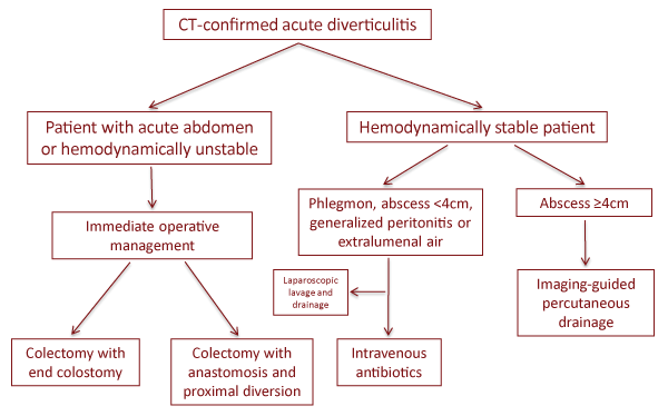
Figure 1: Treatment algorithm for patients with acute diverticulitis
View Figure 1
What is the clinical status of the patient? Patients who present with Systemic Inflammatory Response Syndrome (SIRS) or shock [28] and an acute abdomen require immediate abdominal exploration. This can be performed laparoscopically or open. Most patients with this clinical presentation undergo sigmoidectomy with end colostomy; with the expectation of markedly increased postoperative morbidity and mortality. Currently, many institutions are performing laparoscopic lavage and drainage as a minimally invasive approach with both temporal and curative intents. This therapeutic approach has been mainly reported in case series and institutional experiences and appears to be a safe and effective therapy for selected patients with complicated diverticulitis [29-31]. Randomized controlled trials are currently underway to better evaluate its role in the treatment of complicated diverticulitis [32,33].
The treatment algorithm for patients who are hemodynamically stable at the time of diagnosis of perforated diverticulitis varies according to the physical exam and imaging findings. In this scenario, there is more time to plan the therapeutic approach and to evaluate the response to a given intervention. Patients who are hemodynamically stable with localized peritonitis associated with a phlegmon or evidence of a contained perforation may be managed non-operatively. When an abscess =4cm is present, percutaneous drainage is strongly recommended, especially when patients present with a pronounced systemic inflammatory response [33,34]. Close evaluation is mandated after non-operative treatment, given that 15 to 30 percent of patients have been reported to fail with this approach [35].
Is the perforation free or contained? Diverticular perforation may present as a large perforation with free communication to the peritoneal cavity and fecal peritonitis, or as a micro perforation with mild abdominal pain and a CT showing a phlegmon or small bubbles of free intraperitoneal air.
Large perforations with fecal peritonitis require operative treatment, most commonly a sigmoid colectomy with end colostomy. After sigmoidectomy, if the surgeon feels that the bowel ends are appropriate for reconnection and the peritoneal contamination is limited, primary colorectal anastomosis can be performed. This technique with a diverting loop ileostomy has shown comparable results to sigmoidectomy with end colostomy [36,37]. Gawlick and collegues [36] evaluated 2018 patients who underwent operative treatment for perforated diverticulitis. Of these patients, the majority underwent sigmoidectomy with end colostomy and 17 percent (n=340) underwent resection with anastomosis and diverting loop ileostomy. The two groups were comparable in demographics and disease presentation. Septic patients who underwent sigmoidectomy with end colostomy had significantly more wound infections (14.6% vs. 8.6%, P=0.02), but there were no significant differences in organ-space infection, dehiscence, return to the operating room, postoperative sepsis, or length of hospital stay. Binda et al. [37] randomized 90 patients with perforated diverticulitis and peritonitis to either sigmoidectomy with end colostomy or sigmoidectomy with primary anastomosis and diverting loop ileostomy. Although the trial was closed prematurely because of low accrual of patients, no significant differences were seen in overall postoperative morbidity or mortality. Upon follow up, patients who underwent end colostomy takedown had increased postoperative morbidity compared to those undergoing loop ileostomy reversal (23.5% vs. 4.5%, P=0.058).
Patients who are hemodynamically stable without generalized peritonitis and are found to have a left lower quadrant phlegmon or scattered free air on CT can be managed nonoperatively. In our experience these patients are at a higher risk for developing a diverticular abscess and a repeat CT of the abdomen and pelvis is recommended if signs and symptoms of infection persist.
Summary
Complicated diverticulitis with suspected or confirmed diverticular perforation is becoming an increasingly common disease presentation. However, the pathophysiology, clinical impact, and natural history of diverticular perforation are largely unknown. Theories have been postulated and risk factors have been identified but none completely explain the occurrence of diverticular perforation. Perforated diverticulitis has many clinical presentations that will largely depend on the size of the perforation, degree of peritoneal contamination, and inflammatory response. The majority of patients with perforated diverticulitis can be managed non-operatively with good results. Operative treatment, when necessary, is associated with increased morbidity and mortality. Laparoscopic lavage and colonic resection are minimally invasive approaches that are safe and associated with lower postoperative morbidity and mortality compared to open Hartmann�s procedure and colectomy.
References
-
Hughes LE (1969) Postmortem survey of diverticular disease of the colon. I. Diverticulosis and diverticulitis. Gut 10: 336-344.
-
Parks TG (1969) Natural history of diverticular disease of the colon. A review of 521 cases. Br Med J 4: 639-642.
-
Parks TG (1969) Reappraisal of clinical features of diverticular disease of the colon. Br Med J 4: 642-645.
-
Feingold D, Steele SR, Lee S, Kaiser A, Boushey R, et al. (2014) Practice parameters for the treatment of sigmoid diverticulitis. Dis Colon Rectum 57: 284-294.
-
Morris AM, Regenbogen SE1, Hardiman KM1, Hendren S1 (2014) Sigmoid diverticulitis: a systematic review. JAMA 311: 287-297.
-
American Gastroenterological Association (2001) The Burden of Gastrointestinal Diseases.
-
Munson KD, Hensien MA, Jacob LN, Robinson AM, Liston WA (1996) Diverticulitis. A comprehensive follow-up. Dis Colon Rectum 39: 318-322.
-
Hinchey EJ, Schaal PG, Richards GK (1978) Treatment of perforated diverticular disease of the colon. Adv Surg 12: 85-109.
-
M�kel� J, Kiviniemi H, Laitinen S (2002) Prevalence of perforated sigmoid diverticulitis is increasing. Dis Colon Rectum 45: 955-961.
-
arfwidsson S, Knock Ng, Lehmann L, Winberg T (1964) Pathogenesis of multiple diverticula of the sogmoid colon in diverticular disease. Acta Chir Scand Suppl 63: SUPPL 342:1-68.
-
Sethbhakdi S (1976) Pathogenesis of colonic diverticulitis and diverticulosis. Postgrad Med 60: 76-81.
-
MORSON BC (1963) THE MUSCLE ABNORMALITY IN DIVERTICULAR DISEASE OF THE COLON. Proc R Soc Med 56: 798-800.
-
Fleischner FG (1971) Diverticular disease of the colon. New observations and revised concepts. Gastroenterology 60: 316-324.
-
Fleischner FG, Ming SC (1965) Revised concepts on diverticular disease of the colon: so called diverticulitis, diverticular sigmoiditis and perisigmoiditis; diverticular abscess, fistula and frank peritonitis. Radiology 84:599-609.
-
Hughes LE (1975) Complications of diverticular disease: inflammation, obstruction and bleeding. Clin Gastroenterol 4: 147-170.
-
Tagliacozzo S, Tocchi A (1997) Antimesenteric perforations of the colon during diverticular disease: possible pathogenetic role of ischemia. Dis Colon Rectum 40:1358-1361.
-
Tursi A (2010) Colonic microflora imbalance and diverticular disease. Dig Liver Dis 42: 458.
-
Korzenik JR; NDSG (2008) Diverticulitis: new frontiers for an old country: risk factors and pathogenesis. J Clin Gastroenterol 42: 1128-1129.
-
Jurowich CF, Jellouschek S, Adamus R, Loose R, Kaiser A, et al. (2011) How complicated is complicated diverticulitis?--phlegmonous diverticulitis revisited. Int J Colorectal Dis 26: 1609-1617.
-
von Rahden BH, Kircher S, Thiery S, Landmann D, Jurowich CF, et al. (2011) Association of steroid use with complicated sigmoid diverticulitis: potential role of activated CD68+/CD163+ macrophages. Langenbecks Arch Surg 396: 759-768.
-
Von Rahden BH, Kircher S, Landmann D, Schlegel N, Lazariotou M, et al. (2012) Glucocorticoid-induced tumour necrosis factor receptor expression: a potential molecular link between steroid intake and complicated diverticulitis? Colorectal Dis 14:1276-1286.
-
Mimura T, Bateman AC, Lee RL, Johnson PA, McDonald PJ, et al. (2004) Up-regulation of collagen and tissue inhibitors of matrix metalloproteinase in colonic diverticular disease. Dis Colon Rectum 47: 371-378.
-
Chapman J, Davies M, Wolff B, Dozois E, Tessier D, et al. (2005) Complicated diverticulitis: is it time to rethink the rules? Ann Surg 242: 576-581.
-
Humes DJ, Fleming KM, Spiller RC, West J (2011) Concurrent drug use and the risk of perforated colonic diverticular disease: a population-based case-control study. Gut 60: 219-224.
-
Wilson RG, Smith AN, Macintyre IM (1990) Complications of diverticular disease and non-steroidal anti-inflammatory drugs: a prospective study. Br J Surg 77: 1103-1104.
-
Fuchs HF, Broderick RC, Harnsberger CR, Chang DC, Mclemore EC, et al. (2014) Variation of outcome and charges in operative management for diverticulitis. Surg Endosc.
-
Smith AN, Shepherd J, Eastwood MA (1981) Pressure changes after balloon distension of the colon wall in diverticular disease. Gut 22: 841-844.
-
American College of Chest Physicians/Society of Critical Care Medicine Consensus Conference: definitions for sepsis and organ failure and guidelines for the use of innovative therapies in sepsis (1992) Crit Care Med 20:864-874.
-
Karoui M, Champault A, Pautrat K, Valleur P, Cherqui D, et al. (2009) Laparoscopic peritoneal lavage or primary anastomosis with defunctioning stoma for Hinchey 3 complicated diverticulitis: results of a comparative study. Dis Colon Rectum 52: 609-615.
-
White SI, Frenkiel B, Martin PJ (2010) A ten-year audit of perforated sigmoid diverticulitis: highlighting the outcomes of laparoscopic lavage. Dis Colon Rectum 53: 1537-1541.
-
McDermott FD, Collins D, Heeney A, Winter DC (2014) Minimally invasive and surgical management strategies tailored to the severity of acute diverticulitis. Br J Surg 101: e90-99.
-
Swank HA, Vermeulen J, Lange JF, Mulder IM, van der Hoeven JA, et al. (2010) The ladies trial: laparoscopic peritoneal lavage or resection for purulent peritonitis and Hartmann's procedure or resection with primary anastomosis for purulent or faecal peritonitis in perforated diverticulitis (NTR2037). BMC Surg 10:29.
-
Kumar RR, Kim JT, Haukoos JS, Macias LH, Dixon MR, et al. (2006) Factors affecting the successful management of intra-abdominal abscesses with antibiotics and the need for percutaneous drainage. Dis Colon Rectum 49: 183-189.
-
Brandt D, Gervaz P, Durmishi Y, Platon A, Morel P, et al. (2006) Percutaneous CT scan-guided drainage vs. antibiotherapy alone for Hinchey II diverticulitis: a case-control study. Dis Colon Rectum 49: 1533-1538.
-
Cinat ME, Wilson SE, Din AM (2002) Determinants for successful percutaneous image-guided drainage of intra-abdominal abscess. Arch Surg 137: 845-849.
-
Gawlick U, Nirula R (2012) Resection and primary anastomosis with proximal diversion instead of Hartmann's: evolving the management of diverticulitis using NSQIP data. J Trauma Acute Care Surg 72: 807-814.
-
Binda GA, Karas JR, Serventi A, Sokmen S, Amato A, et al. (2012) Study Group on Diverticulitis. Primary anastomosis vs nonrestorative resection for perforated diverticulitis with peritonitis: a prematurely terminated randomized controlled trial. Colorectal Dis 14:1403-1410.
