International Journal of Sports and Exercise Medicine
Influence of 120-Day Stimulated Microgravity with Countermeasures on Human Muscle Musculo-Tendinous Stiffness and Contractile Properties
Yuri A Koryak*
SSC- Institute of Biomedical Problems RAS, Russia
*Corresponding author: Yuri Koryak, SSC - Institute of Biomedical Problems RAS, 76-A Khoroshevskoye Shosse 123007 Moscow, Russia, E-Mail: yurikoryak@mail.ru
Int J Sports Exerc Med, IJSEM-1-006, (Volume 1, Issue 2), Research Article; ISSN: 2469-5718
Received: March 23, 2015 | Accepted: May 12, 2015 | Published: May 15, 2015
Citation: Koryak YA (2015) Influence of 120-Day Stimulated Microgravity with Countermeasures on Human Muscle Musculo-Tendinous Stiffness and Contractile Properties. Int J Sports Exerc Med 1:006. 10.23937/2469-5718/1510006
Copyright: © 2015 Koryak YA. This is an open-access article distributed under the terms of the Creative Commons Attribution License, which permits unrestricted use, distribution, and reproduction in any medium, provided the original author and source are credited.
Abstract
The effect of long-term (120-day) 5° head-down tilt (HDT) bed rest with countermeasures (physical training - PT) on the contractile properties and musculo-tendinous stiffness (MTS) of the human triceps surae muscle (TS) was studied in four healthy young women subjects. The training sessions were conducted for 60 min each day for 6-days a week for 14 weeks, and 30-40 min twice a day for 2 weeks under the experiment conditions. The maximal voluntary contraction (MVC), and isometric twitch contraction (Pt), and electrically evoked tetanic tension at 150 impulses s-1 (Po), time-to-peak tension (TPT), and half relaxation time (1/2 RT), and total contraction time (TCT) of the twitch were determined. The difference between Po and MVC expressed as a percentage of Po and referred to a force deficiency (FD). The MTS was determined according to the electromechanical delay (EMD) value during the explosive voluntary contraction. Surface electrodes sensed electromyographic activity (EMG) in the soleus muscle. A separate timer was used to determine total reaction time (TRT). Pre-motor time (PMT) was taken to be the time interval from the delivery of the signal to change in EMG. Electromechanical delay (EMD) was the time interval between the change in EMG and movement muscle force production. After HDT with PT the TPT, 1/2 RT and TCT of the TS decreased by 4%, 17%, and 19%, respectively. The MVC, Pt, and Po decreased by 3% (p>0.05), and 14%, and 9%, respectively. The FD had decreased significantly by 10 % (p< 0.02). The rate of increase of electrically evoked tetanic did not change significantly during HDT with PT, but the rate of increase in isometric voluntary tension development was increased. The EMD, PMT, and TRT decreased by 12.2% (p< 0.05), 5.3% and 7.3% (p< 0.01), respectively. The results obtained demonstrate that PT counteracted the negative effect of mechanical unloading, but the total physical load (mainly its intensity) was obviously insufficient to prevent changes to the mechanical properties of the muscular system, although both a nervous and muscular response to PT was observed.
Keywords
Bed rest, Physical training, Triceps surae muscle, Electromechanical delay, Musculo-tendinous stiffness, Contractile properties
Introduction
It is well known that the unloading of the musculoskeletal system by actual or simulated microgravity causes numerous changes in the musculoskeletal system, such as muscular atrophy and decreased contraction strength, both after relatively short-term (10-17) [1-6] and long-term (>5 weeks) periods of unloading [5,7-12]. The deterioration of musculoskeletal function causes no direct health hazards and does not affect the capacity for work during short-term space missions. However, during long-term missions, serious problems can arise after returning back to Earth, unless the negative effects of microgravity are mitigated. Therefore, measures to prevent the complete adaptation of the human body to microgravity conditions are vital, as well as supporting the effective functioning of all body's systems, which have adapted to life in the terrestrial gravitational field through phylogenesis and ontogenesis.
Physical exercise (physical training - PT) has been proposed as a potential countermeasure (method) to microgravity-induced effects on the size (mass) and function of skeletal muscles [13,14]. High-resistance physical exercises are known to be effective in increasing the size (or cross-sectional area) or contractile force of muscles [15,16]. Therefore, high-intensity resistance training may be successfully applied during unloading in order to counteract the deterioration of the contractile functions of the muscle.
Surface electromyogram (EMG) data show the electrical activity of muscle and have been used in the analysis of human movement. The electromechanical delay (EMD) is traditionally defined as the time lag between the onset of muscle electrical activation and the onset of force production [17]. During this time frame, a sequence of several physiological events transduces the electrical processes of muscle activation into a mechanical phenomenon: (i) the propagation of the action potential along the sarcolemma and the T-tubule system; (ii) the coupling between the dihydropyridine and ryanodine receptors and the following release of Ca2+ from the sarcoplasmic reticulum; (iii) the interaction among Ca2+, troponin and actin; (iv) the cross-bridges formation; and (v) the myosin heads rotation with subsequent force transmission to the tendon insertion point through the elongation of the series elastic components (SEC). Muscle force is registered only when the contractile elements of the muscle shorten, thus stretching the SEC, which participates in the transmission of force to tendons and joints [18,19].
Because of the several approaches with different experimental protocols, EMD values from different human skeletal muscles have been found to vary between 8 and 127 ms [20-23]. Changes in any of the previously listed events could potentially induce EMD alterations [24-30].
If the contraction strength changes, significant changes are seen in the EMD values and, therefore, in the SEC [31-33]. This confirms the idea that the time until the SEC stretches is a primary EMD determinant. Furthermore, the SEC structures, originally consisting of an active part (located in the myofibrilare) and a passive one (represented mainly by the aponeurosis and tendon) [34], may contribute to the EMD value.
EMD includes the time related to electromechanical muscle excitation, cross-bridge activation and stretching of the SEC [35]. The time required to stretch the elastic components of the musculo-articular system [36,37] is the prevailing EMD component and thus a measure of the series elastic stiffness.
Musculo-tendinous stiffness (MTS) is known to increase after of strength training [38-40], and it may therefore be expected that the EMD would be shorter in those subjects who practiced PT in microgravity conditions, compared to those who were not subject to PT [41].
The EMD changes can be attributed, first of all, to changes in muscle SEC stiffness. Muscle stiffness is described as the ratio between the force and length of stretching. A mechanically stiff muscle will transmit more force if the SEC stretches a small amount and, on the contrary, mechanically "gentle" (or "slack") tissue requires a significantly stronger muscle contraction in order to stretch the SEC and produce measurable force. "Slack" tissues require more time from the moment of activation to the start of force generation, i.e. their EMD is longer and it is shortened when muscle stiffness is increased before muscle tension [31,35,42-44]. Therefore, the EMD may become a criterion for the differences in stiffness of the musculo-tendinous complex (MTC) under different conditions.
Unloading leads not only to the loss of muscle mass and muscle dysfunction (which seems to be clinically relevant), but also affects other functionally important structures of the musculoskeletal system, which undergo changes under the influence of microgravity (in particular, the tendon serially connected to the muscle). It is already known that the tendon not only provides structural connections between a muscle and a bone, but also plays the main role in the transmission of force produced by contractile muscle elements to the skeleton. Therefore, the tendon can change the length, and, therefore, the force of the contractile elements connected serially depends on the degree of variation of tensile strength [34,35]. The degree to which a tendon deforms in response to a muscle contraction depends on the mechanical properties of the tendon [46-48], since the tendon is not an inert structure and like a muscle it can change with the physical load level. The mechanical properties of the tendon may be adapted to changes in load level [49-52]. For example, a higher load is associated with tendon hypertrophy [51]. Therefore, if unloading causes decreased stiffness in the muscle MTC, then it is vital to understand whether load (physical training) can prevent the potentially harmful effect of unloading.
Firstly, the objective of this study is to evaluate the effect of PT on the contractile properties of the TS in a group of young women in order to reduce the negative effects of mechanical unloading caused by long-term bed rest, and secondly, to assess the degree of EMD changes in the subjects of the study undergoing a strict bed rest regimen but also engaging in PT. We suppose that if the decreased MTC stiffness is caused by unloading, then PT during bed rest may prevent the detraining of skeletal muscles and reduce the negative effect of unloading.
Materials and Methods
The study was approved by the local Institute Ethical Committee and had been performed in accordance with the principles of the 1975 Declaration of Helsinki on the use of human subjects in experiments.
Participants
A group (n=4) of healthy female volunteers aged 28.0 ± 1.1 years (range 26 - 31), 162.3 ± 4.2 cm in height (range 174 - 155), and mass 59.9 ± 2.3 kg (range 53 - 62.2) participated in the study. During preliminary visits to the laboratory, the subjects were informed of the objectives and methods of the study and were introduced to all the procedures for evaluating voluntary muscle contractions and those induced by electricity. After that, each subject signed an Informed Consent Form for participating in the experiment.
Selection of subjects were based on an evaluation that consisted of taking a detailed medical history, complete blood count, urinalysis, resting electrocardiogram, and a selection of blood chemistry analyses, which included the estimation of concentrations of fasting blood glucose, bload urea nitrogen, creatinine, lactic dehydrogenase, serum transaminase bilirubin, uric acid, and cholesterol, as well as an evaluation of their physical state using a bicycle ergometer stress-test [1]. No subject was taking medication at the time of the study, and all the subjects were non-smokers and recreationally active but not especially well trained.
Bed rest
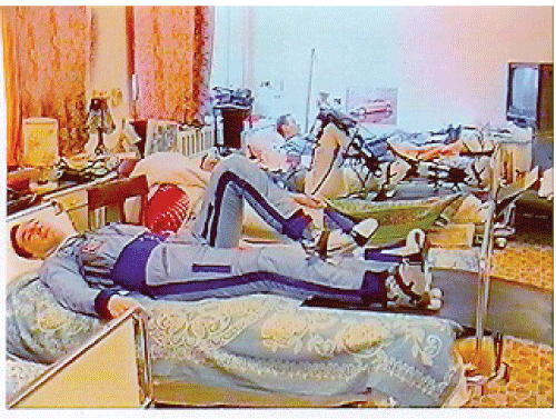
Bed rest during 120-days in an antiorthostatic position (5° HDT) of the body was used as a model of the long-term hypokinesia/hypodynamia effect of spaceflight (Figure 1). The 5° HDT position was chosen since various physiological alterations induced by actual spaceflight [53].
.
Figure 1: The position subject of the test bed rest. A participant performing the exercise in the 5 head-down tilt position. A participant performing the exercise in the 5° head-down tilt position.
View Figure 1
During this 120-day experiment, the subjects were housed 24 h day-1 in the Human Research Facility of the Health Ministry Institute of Biomedical Problems. During bed rest, subjects remained in the HDT position continuously for all activities including excretory functions, showering, eating and PT.
Exercise regimens
Details of the training program and performance tests have been provided elsewhere [54,55]. Running on a treadmill in an upright position was the main training procedure. The subjects of the study were taught to do dynamic physical exercises in the recumbent position, lying on their backs, using a specialized system to pull themselves to the rubber track of the treadmill (the load degree varied from 0 to 70 kg). The study subjects started PT 12 16 days after the beginning of bed rest.
PT used during long-term spaceflights in the Russian space station "MIR" included a warm-up (walking on a treadmill for 5 min), and low (2 min), moderate (2 min) and maximum (1 min) intensity running. PT was scheduled over a 4-day cycle: 3-days of training, 1-day of rest. The training sessions were conducted for 60 min each day for 6-days a week for 14 weeks, and 30 40 min twice a day for 2 weeks under the experiment conditions. In addition, taking into consideration the anatomical and physiological specificities of a woman's body, the total physical load was reduced to 70 % of that usually exposed to by men, and when the expanders were used for muscle-strengthening exercises; the load was reduced by 25-30%.
Experimental protocol and measurements
The experiment procedure and the set-up used to record the voluntary and electrical (involuntary) evoked contractions of the TS were the same as has been previously described [56,57]. In brief, the subject was seated comfortably on a special chair in a standard position (at a knee joint angle between the tibia and the sole of the foot of proximately 90°). The position of the seat was adjusted for the individual and then firmly secured. A rigid leg position provided the isometric regimen of the muscle contraction. The dynamometer (maximal force 500N, sensibility 0.5N) was a steel ring with a saddle-shaped special block attached to the Achilles tendon. The resting pressure between the force transducer and the tendon was constant for all the subjects and was set at 5kg. The dynamometer was calibrated after each test by loading it with various weights.
The contractile properties of the TS were tested twice: 10-8 days before the beginning of the bed-rest and after it ended. The test protocol was identical for both prebed rest and postbed rest.
Tension properties
The contractile properties of the TS were estimated according to the mechanical parameters of a voluntary and electrically evoked contraction (maximal isometric twitch and tetanic contractions, double stimulation).
The subjects were instructed to respond to an auditory signal by exerting maximal force. From 2-3 maximal contraction were usually recorded from each subject until maximal force concentrations was obtained. There was a 1-min rest between the sets. The MVC was determined as the highest value of voluntary force recorded during the entire contraction.
Maximal isometric twitch contraction, and tetanic contraction, and double contraction, of the TS were induced by the electric stimulation of the tibial nerve by an electrical stimulator (Neuromuscular Stimulator Universal "NSU-1", USSR). Electrical stimulation was applied through monopolar electrodes, one (the cathode) 1 cm in diameter, was located in the popliteal fosse (tibial nerve) which is the site of lowest resistance, and the other electrode (the anode) was positioned on the lower one-third of the front surface of the femur. Voltage was increased in a stepwise manner until maximal twitch responses were evoked. A single stimulus was given every 30 s. From the maximal isometric twitch responses the force (Pt) were measured [5,56].
Myoelectrical signals or surface action potentials (M-wave) were recorded using two-size electrodes (Ag-AgCl, 8mm contact diameter, 25mm inter-electrode distance). These active electrodes were applied over the lateral side of the soleus muscle approximately 15 cm proximal from the calcaneous. The skin area under the electrodes was cleaned carefully with ethyl alcohol and gently abraded with a special abrasive to achieve inter-electrode impedance below 2,000 Ω. The ground electrode (Ag-AgCl, 7.5 × 6.5cm) was placed in the proximal part of the ankle between the pickup and stimulating electrodes. The peak-to-peak amplitudes of the M-waves were measured as has been described by Sica and McComas [58].
The maximal force (Po) was evoked by delivering supramaximal voltage electrical rectangular pulses of 1ms at a frequency of 150 impulses/s-1 [5,6,41,56]. The total duration of the tetanic stimulation was equal to approximately 0.5 s. The difference between Po and MVC was expressed as a percentage of the Po value and referred to as force deficiency (Pd) as has been calculated before [41,56]. This parameter reflects the capability of a certain part of the motor pool [56].
After a rest of 5 min the motor nerve was stimulated at various intervals. Supramaximal double stimuli at 3, 4, 5, 10, 20, and 50ms intervals after the first one were applied [5,6,56]. The maximal strength (amplitude) of the muscle contraction was determined and expressed as a percentage of the twitch contraction.
Velocity properties
The time from the moment of stimulation to peak twitch (TPT), the time from contraction peak to half-relaxation (1/2 RT) and total contraction time (TCT) - the time from the moment of stimulation to the total muscle relaxation were calculated by the tendogram of isometric twitch [41,56]. The accuracy of measurement was 1ms.
The tetanic index (TI) was estimated using the Pt / Po amplitude ratio [5,56].
Force-velocity properties
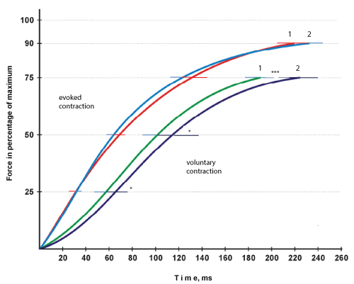
The rate of development of increased muscle tension was calculated from the tendogram isometric voluntary contraction after the instruction to exert the fastest and strongest contraction using a relative scale, i.e. the time of reaching 25%, 50%, 75%, and 90% of maximum tension [56]. Voluntary TS contraction in response to a light stimulus was taken as an explosive ballistic muscle movement.
Similarly, the rate of rise of electrically evoked contraction in response to electric stimulation of the tibialis nerve with a frequency of 150 impulses s-1 was determined [5]. The accuracy of measurement was 1ms.
The maximum rate (dPo / dt) of the rise of tension was additionally calculated from the force-time curve of the isometric voluntary contraction by the differentiation of the analogue signal. Similarly, the maximum rate of the electrically evoked contraction (dPo / dt) was calculated.
Electromechanical delay
The subjects were informed to extend the foot as quickly and as strongly as possible after the visual signal lamp (∅ 7mm, 1W) - was placed at eye level 1m in front of the subject signal (Figure 2). The signal was given from an electrical timing unit. After cessation of the signals the subject was instructed to relax his muscles as quickly as possible. The signal duration was 2.5 s and intervals between signals were set in a random manner and ranged between 1.4 to 4.0 s.
.
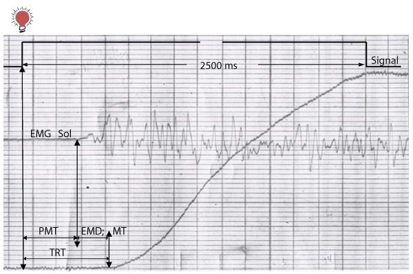
Figure 2: Schematic presentation of a sample showing total reaction time (TRT) with its premotor (PMT) and motor time (MT) or electromechanical delay (EMD) and electromyogram (EMG) of the soleus (Sol) muscle.
View Figure 2
A specialized timer was used which recorded the development of the TS mechanical response simultaneously with the light signal for the beginning of the movement. The TRT was estimated from the mechanogram; it was calculated as the interval between the light signal and the beginning of the contraction. The TRT was represented by the PMT which is defined as the interval between the presentation of the light stimulus to the initial changes in the electrical activity of the muscle and by the motor time (MT, or electromechanical delay, EMD), which is defined as the interval from the moment electric activity of the agonist muscle is detected to the beginning of movement, i.e. to the moment of contraction development [59] (Figure 2). The thresholds for EMGs and force were 36μV and 5 N, respectively.
Each study subject performed three attempts with a rest interval of at least 1 min, and the best attempt was used to estimate the TRT, PMT, and EMD.
The results of the experiment were simultaneously recorded on magnetic tape and was also recorded on a storage oscilloscope.
Statistics
Conventional statistical methods were used for the calculation of means and standard errors of the mean. Differences between baseline (background) values of the subject and those post exposure (bed- rest) were tested for significance by Student's paired t test. Values are given as means ± S.E.M. throughout. Significant differences between means were set at the p< 0.05 level. The percentage changes for pre- and postbed-rest were calculated.
Results
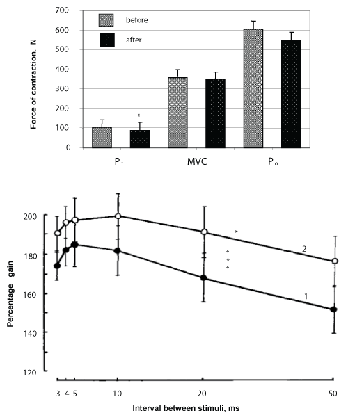
The effects of 120-day HDT with PT on contractile properties the TS are illustrated by Figure 3, top panel. The analysis of the results obtained revealed a not significantly decrease in the tension properties of the muscle. Thus, isometric Pt decreased by 13.6% [pre 101.0 (SEM 17.6) N compared to post 87.3 (SEM 6.8) N; p< 0.05], MVC by 3.1% [pre 52.2 (SEM 55.9) N compared to post 341.4 (SEM 25.5) N; p>0.05] and Po by 9.4 % [pre 594.5 (SEM 66.7) N compared to post 538.6 (SEM 25.5) N; p>0.05]. The Pd decreased significantly by 10.0% [pre 40.2 (SEM 8.2)% compared to post 36.2 (SEM 5.5)%; p< 0.05].
.
Figure 3: The effect of a 120-day 5° head-down tilt (HDT) with physical training on maximal voluntary contraction (MVC), and evoked tetanic tension at 150 Hz (Po), and at maximal twitch tension (Pt) (top panel) and on the maximal force contraction of triceps surae muscle in response to supramaximal twin stimuli at 3, 4, 5, 10, 20, and 50ms (bottom panel) 1 - before 2 - after
*-p< 0.05; **** -p< 0.001
View Figure 3
Mean changes in isometric strength of the TS under paired stimulation of maximal intensity at interpulse intervals of 3, 4, 5, 10, 20, and 50ms can be seen in Figure 3, low panel. As may be seen the greatest force of contraction was observed at intervals of 4 10ms and decreases or increases outside this range was accompanied by considerable decreases (p< 0.05) but with similar trends of tension developed by the muscle. There were differences in the curve at identical interpulse intervals the relative increase in force of contraction after 120-day HDT with PT being significantly greater by comparison with the initial values (p< 0.001).
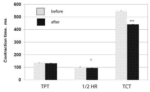
The change of mean time of isometric twitch contraction for the TS after the 120-day HDT effect with PT is given in Figure 4. As is seen from the analysis of the data, exposure to HDT conditions was accompanied by a statistically significant increased muscle contraction and increased relaxation velocity. Thus, TPT decreased by a mean of 3.5% [pre 135.5 (SEM 11.7) ms compared to post 130.8 (SEM 6.0) ms; p>0.05], 1/2 RT and TCT decreased by 7.4% [pre 101.5 (SEM 10.0) ms compared to post 94.0 (SEM 10.2) ms; p< 0.05] and 19.3 % [pre 546.8 (SEM 25.3) ms compared to post 441.3 (SEM 19.8) ms; p< 0.001], respectively.
.
Figure 4: The effect of a 120-day 5° head-down tilt (HDT) with physical training on the isometric twitch time-to-peak tension (TPT), half-relaxation time (1/2 RT), and total contraction time (TCT) of the triceps surae muscle.
*-p< 0.05; **** -p< 0.001
View Figure 4
TI was reduced by 15.8% (p< 0.05).
Changes in the development rate of isometric tension in the TS are given in Figure 5. Analysis of the data provided evidence of an increase in the rate of rise of development of isometric voluntary tension in the TS (p< 0.05). This may be seen as an increase in convexity of the force-time curve estimated according to a relative scale. However, no substantial changes were observed in the electrically evoked isometric tetanic development (p>0.05).
.
Figure 5: The effect of a 120-day 6° head-down tilt (HDT) with physical training on the maximal rates of development of voluntary isometric force and of electrically evoked tetanus. 1 - before 2- after
*- p<0.05; **** -p<0.001
View Figure 5
The analysis of the maximum normalized ratio dPo/dt revealed a not significantly decrease by a mean of 8.4 % [pre 0.83%/ms compared to post 0.78%/ms; p< 0.001]. The analysis of the development of electrically evoked contractions demonstrated not significantly differences throughout the whole period of the development of isometric tension (p>0.05), but at the same time, a minor (by a mean of 14.5%) increase of the maximum Po/dt, compared to the initial one [pre 1.17%/ms compared to post 1.34%/ms; p< 0.001].
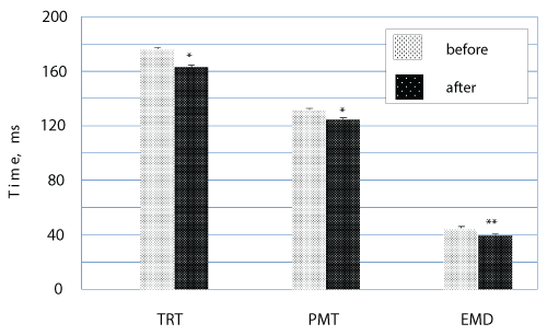
The EMD was significantly higher in explosive voluntary the TS contractions in response to the light signal (by a mean of 12.2%) after 120-day HDT with PT compared to the baseline value [pre 44.9 (SEM 2.0ms compared to post 39.4 (SEM 3.1) ms; p< 0.05] (Figure 6). After mechanical unloading, the PMT decreased by 5.3% (pre 131.8 ± 6.4ms compared to post 124.8 ± 7.1ms; p< 0.05) and the TS decreased by a mean of 7.3% [176.2 (SEM 6.7) ms compared to post 163.4 (SEM 8.3) ms; p< 0.01).
.
Figure 6: The effect of a 120-day 6° head-down tilt (HDT) with physical training on the total reaction time (TRT), premotor (PMT) and electromechanical delay (EMD)
*-p< 0.05; ** -p< 0.01
View Figure 6
Discussion
For the first time, this work shows the effect of chronic unloading on the mechanical properties of the foot extensors in a group of young adult women who have practiced PT and evaluates the program of PT intended to preserve muscle function during periods of unloading. The TS was the muscle tested in the study, since it has been demonstrated that foot extensors were the muscles most affected by unloading, compared to other extensors of the lower limbs [60].
This study considers the program of physical exercises carried out by the study subjects during 120-days of strict HDT, while the International Space Station team members carry out a similar program over 6 months. We studied the advantages of this program in order to protect various contractile elements of the skeletal muscle. This study may be considered unique due to the duration of the unloading period (120-days) with the use of women as study subjects and the examination of the physical adaptation of the muscular system to longer periods of unloading.
Our results indicate that the 120-day of simulated microgravity with PT is associated with a not significant decrease in the contractile properties of the TS. For instance, the MVC after the 120-day HDT with PT decreased by 3%, the TCT decreased by 4%, and the rising curve of isometric voluntary contraction underwent fewer changes, which, in general, confirms the importance of PT in the preservation of muscle function , especially under conditions of mechanical unloading. However, the physical training program applied, as well as the regimen of exercises carried out, did not completely prevent the negative effect of unloading on TS function.
Less significant changes in the MVC in response to unloading may mean that PT contributes not so much to the relative preservation of functions, as to the increase in the role of central nervous regulation, which is the main factor determining the MVC. The latter is confirmed by fewer changes in the Pd, which is indicative of the increase of the central drive in the motor control of voluntary movements by the nervous system. These results comply with previously obtained data that PT contributes to increased synchronization and activation of motor units [61] and permits us to suppose that nervous adaptation may take place and it significantly contributes to the increase in the MVC which was observed during the 120-day HDT with PT and decrease with HDT without countermeasures [62,63]. Therefore, PT counteracts the muscle mass loss related to inactivity and prevents changes in the composition of muscle fibers caused by non-use [64]. This complies with previously obtained data that contractile adaptation to training occurs independently of changes in fiber types [65]. Since we were not able quantitatively to assess the intensity of the exercises performed and the device applied had certain limitations, this could have led to the study subjects performing a greater volume of exercises, but with less intensity. Data on the use of high resistance exercises with high intensity confirms their importance in the protection of muscle function from atrophy. Thus, balanced PT consisting of high resistance and intense exercises (2-3 days/week) and aerobic exercises (~ 4 days/week) is more effective in the preservation of muscle function during prolonged bed rest (Trappe et al. 2007, 2008, 2009). Moreover, training with highly intensive resistance exercises lasting for ~ 7min/week during the bed rest period was more effective when compared to exercises lasting for more than 60min/week under actual zero gravity conditions [66].
We expected that changes in the Pt and Po would correspond to MVC changes, namely, to their increase with PT [14] and decrease during bed rest without PT [14]. However, in this study, Po even slightly decreased (~ 9%) after PT, which led to a tendency toward a decrease in the Pt / Po after the 120-day HDT with PT being observed. Changes in the Pt / Po may reflect the degree of changes in the muscle tensile strength. The literature indicates that disuse induced an increase in muscle and joint stiffness and a decrease in the range of motion [11,67]. Decreased muscle tensile strength permits the body to transmit the tension developed by sarcomeres of the contractile tissue more effectively and thus the Pt / Po increases. On the contrary, increased muscle tensile strength during PT may result in a decreased Pt / Po [68]. We suppose that in our experiment PT during the HDT contributed to the increased muscle tensile strength to a certain extent. Training may lead to a decreased Pt by causing an increase in the muscle tensile strength, which is described in this work.
Within the framework of this study, PT was performed every third day, which only allowed the subjects to carry out two sessions a week. Furthermore, taking into consideration the fact that physical training was performed twice a week by the end of the study but total physical load was reduced down to 70% of that usually applied to men (see the Methods), then it seems that the total volume and, especially, the intensity of exercises, was insufficient to support the mechanical properties of the TS tendon. Furthermore, PT significantly reduced the level of decrease in MTC stiffness when compared to unloading without training [41]. It should be noted that the tendon is not an inert structure, and a skeletal muscle also changes with the level of physical load; in other words, the mechanical properties of the tendon may adapt to load changes.
During gravity loading (0 G), the human TS tendon is exposed to very high rhythmic load due to constant muscle use and the transmission of muscle force related to heel activity (heel-and-toe movements), which require stabilization and movement of the body during movement (walking and running) [69]. Therefore, during unloading the full volume of exercises (load level, frequency and duration) should exceed the threshold level in order to prevent changes to the mechanical properties of the tendon completely. The walking load produced by the Achilles tendon is ~ 210 kg/cm2 [70], i.e. for a person weighing 70 kg, the lifting and dropping of body weight in 1g would lead to the development of tension in the Achilles tendon equal to ~ 200kg/cm2. Therefore, if we suppose that ordinary walking is a stimulus sufficient to maintain the mechanical properties of the tendon under normal gravity conditions, then the threshold PT level required for the prevention of any deterioration during unloading should be more than or equal to the body weight. Hence, it may be supposed that although the load level applied in this study has been sufficient, the training regimen followed during unloading has been significantly lower than the load frequency during ordinary walking. Therefore, the required load frequency should obviously be higher in order to exceed the threshold volume. The latter is confirmed by a study when during 20 days of bed rest, PT was not only performed every day (16 out of 20 days), but also with load/weights approaching the subjects body weight [65]. Moreover, the analysis of exercises used in the training process within the frames of this study revealed a lack of exercise for training the foot extensors. And if we take into consideration the fact that the total PT load was reduced (see the "Methods"), it may be the main (or even principal) cause of incomplete preservation of muscle function during unloading.
PT and disuse have often been associated with muscle hypertrophy and atrophy, respectively [64]. In the present experiment no direct measurement of the TS mass muscle was made; however, the peak-to-peak amplitude of the maximal M-wave is an indirect measurement of the excitable muscle mass. The M-wave amplitude did not change significantly with either PT or HDT [10.4 (SEM 0.9)mV compared to post 11.0 (SEM 1.0)mV], suggesting that little alteration in muscle mass occurred.
During many everyday activities, the ability to produce high muscle force per time unit is more important than the ability to generate high force. The rate of force development depends on many factors, in particular, on the duration of the excitation-contraction process, on the force-velocity properties of muscle fibers (even during isometric contraction due to tendon structure deformity) and SEC stiffness. Therefore, decreased MTC stiffness may be the reason for the decreased rate of development of TS contraction after unloading, since SEC stiffness in known to be an important factor determining the rate of muscle force development [71]. In other words, as indicated in the Introduction, the time of SEC stretching by contractile elements determines the EMD value [18]. Therefore, changes in MTC (SEC) stiffness after hyperactivity/hypoactivity mainly explain changes in EMD. In addition, all the structures of the SEC, classically composed of an active part (located in myofibrils) and a passive part (mainly aponeurosis and tendon) [34], could contribute differently to EMD. Nordez et al. [22] used the noninvasive methodology (a real-time ultrasonography) determine the relative contribution of the passive part of the SEC (47.5 ± 6.0% of EMD) and each of the two main structures of this component (aponeurosis and tendon, representing 20.3 ± 10.7% and 27.6 ± 11.4% of EMD, respectively).
This study demonstrated that the rate of isometric voluntary contraction development performed after the instruction to exert the fastest contraction had decreased after unloading, which confirms the theoretical relationship between the decreased muscle MTC stiffness and decreased rate of transmission of the contractile force and therefore the rate of contraction development, but to a significantly less extent when compared to similar conditions without PT (Figure 3). Our results in the present study indicate that PT during bed rest results in an increase in MTS (MTC), whereas a lack of training is associated with a decrease of this parameter [40]. Therefore, PT decreases the EMD under muscular system unloading conditions.
The changes in elastic properties due to PT have been well documented. Endurance training resulted in an increase in the SEC stiffness in the soleus muscle of rats, associated with an increase in type I fibres [72]. Both jump and endurance training also appear to increase both collagen concentration [73,74] and muscle passive stiffness. The soleus rat muscles submitted to plyometric training had faster twitch fibres and a lower SEC stiffness than controls [75-78] reported that human subjects given 8 weeks of maximal effort stretch-shortening cycle exercise training tended to have an increase in the proportion of type II fibres in their vastus lateralis muscles. Kubo et al. [10] reported that the MTS is greater in long distance runners than in untrained individuals.
The results obtained correlate well with data on the decreased EMD after isometric training under bed-rest conditions [39]. The EMD changes during training are mainly attributed to changes in the tendon structure, but it should be noted that tendon stiffness is always increased during any physical activity, both during endurance (Buchanan and Marsh 2001) and isometric training [10,39]. Furthermore, EMD changes correlate very closely with muscle MTC and especially with the active SEC fraction during training [74,76,78].
This study demonstrated that the EMD is sensitive enough and may serve as an indirect marker for measurements of muscle MTC stiffness in order to determine the chronic adaptation of the musculo-tendinous structure of the muscle to the mechanical unloading during PT under real or simulated microgravity conditions. The data obtained demonstrates that a 120-day of simulated microgravity led to decrease the TS mechanical properties and although the adverse effects were reduced thanks to PT, they were not completely prevented by the training program. This permits us to suppose that the volume of exercises and especially the intensity of exercises performed did not exceed the threshold level required for the complete prevention of changes in mechanical properties.
The mechanical muscle responses obtained and the list of physical exercises received from the study subjects, as well as from members of long-term (6-month) space missions confirm the importance of PT in preserving muscle function and the capacity to work during long-term stays under microgravity conditions. Within the framework of this study, the PT program contained mainly low intensity exercises. The inclusion of exercises with higher load and higher intensity into the training process would contribute to a more effective exercise program for the training of skeletal muscles and it would reduce the total training time under zero gravity conditions. In general, PT under microgravity conditions allows subjects to create an increased functional reserve and reduces the effect of unloading observed under real microgravity conditions.
Acknowledgements
The author wishes to express his appreciation to all who contributed to the success of the experiment. He is especially grateful to for his guidance as the IBMP representative of the Exercise Countermeasures Project and the exercise testing group, including I. Amelin, N. Kharitonov, and N. Serikov.
The author gratefully acknowledges the eight women subjects that endured the 120-day confinement.
This work was supported by the Fonds Institute of Biomedical Problems.
References
-
Kozlovskaia IB, Grigor'eva LS, Gevlich GI (1984) Comparative analysis of the effect of weightlessness and its model on the velocity-strength properties and tonus of human skeletal muscles. Kosm Biol Aviakosm Med 18: 22-26.
-
Berg HE, Tesch PA (1996) Changes in muscle function in response to 10 days of lower limb unloading in humans. Acta Physiol Scand 157: 63-70.
-
Narici M, Kayser B, Barattini P, Cerretelli P (2003) Effects of 17-day spaceflight on electrically evoked torque and cross-sectional area of the human triceps surae. Eur J Appl Physiol 90: 275-282.
-
Koryak Yu.A (2008) Neuromuscular changes under the effect of 7 day mechanical unloading of the human muscular system. Fundamental Studies 9: 8-21.
-
Koryak Yu (2011a) The adaptation of human skeletal muscles to load changes. Experimental study. Lap Lambert Academic Publishing GmbH & Co, KG, Germany.
-
Koryak Yu (2011b) Neuromuscular adaptation to short-term and long space flights of the man. In: Grigorev A, Ushakov I (ed) International Space Station. Russia Segment. M. SSC-IMBP RAS 2: 93-123.
-
Hather BM, Adams GR, Tesch PA, Dudley GA (1992) Skeletal muscle responses to lower limb suspension in humans. J Appl Physiol 72: 1493-1498.
-
Berg HE, Larsson L, Tesch PA (1997) Lower limb skeletal muscle function after 6 wk of bed rest. J Appl Physiol (1985) 82: 182-188.
-
LeBlanc A, Lin C, Shackelford L, Sinitsyn V, Evans H, et al. (2000) Muscle volume, MRI relaxation times (T2), and body composition after spaceflight. J Appl Physiol 89: 2158-2164.
-
Kubo K, Kanehisa H, Kawakami Y, Fukunaga T (2000) Elastic properties of muscle-tendon complex in long-distance runners. Eur J Appl Physiol 81: 181-187.
-
Lambertz D, Mora I, Grosset JF, Perot C (2003) Evaluation of musculotendinous stiffness in prepubertal children and adults, taking into account muscle activity. J Appl Physiol 95: 64-72.
-
Koryak Yu, Gidzenko Yu, Shuttlerworth M, Zaletin S, Lonchakov Yu, et al. (2007) Functional properties of the neuromuscular system and their changes after a 7-day spaceflight in the International Space Station. Achivements in Modern Natural Sci 12: 149-150.
-
Nicogossian AE (1982) Countermeasures to space deconditioning. In: Space physiology and medicine. Nicogossian AE, Huntoon C, Pool SL (ed). Lea and Febiger. Philadelphia: 294-311.
-
Koryak Y (1998) Effect of 120 days of bed-rest with and without countermeasures on the mechanical properties of the triceps surae muscle in young women. Eur J Appl Physiol Occup Physiol 78: 128-135.
-
Tesch PA (1991) Training for bodybuilding. In: The encyclopedia of sports medicine. Strength and power in sports. Komi PA (ed). Blackwell Oxford: 370-380.
-
Koriak IuA (1993) Functional properties of the neuromuscular system in athletes of various specialties. Fiziol Cheloveka 19: 95-104.
-
Cavanagh PR, Komi PV (1979) Electromechanical delay in human skeletal muscle under concentric and eccentric contractions. Eur J Appl Physiol Occup Physiol 42: 159-163.
-
Hill AV (1938) The heat of shortening and the dynamic constants of muscle. Proc Royal Soc Lond Ser B Containing Papers of a Biol Character 126: 136-195.
-
Hof AL, Van den Berg J (1981) EMG to force processing I: An electrical analogue of the Hill muscle model. J Biomech 14: 747-758.
-
Hopkins JT, Feland JB, Hunter I (2007) A comparison of voluntary and involuntary measures of electromechanical delay. Int J Neurosci 117: 597-604.
-
Grosset JF, Piscione J, Lambertz D, Pérot C (2009) Paired changes in electromechanical delay and musculo-tendinous stiffness after endurance or plyometric training. Eur J Appl Physiol 105: 131-139.
-
Nordez A, Gallot T, Catheline S, Guével A, Cornu C, et al. (2009) Electromechanical delay revisited using very high frame rate ultrasound. J Appl Physiol 106: 1970-1975.
-
Yavuz SU, Sendemir-Urkmez A, Türker KS (2010) Effect of gender, age, fatigue and contraction level on electromechanical delay. Clin Neurophysiol 121: 1700-1706.
-
Viitasalo JT, Komi PV (1981) Interrelationships between electromyographic, mechanical, muscle structure and reflex time measurements in man. Acta Physiol Scand 111: 97-103.
-
Granata KP, Ikeda AJ, Abel MF (2000) Electromechanical delay and reflex response in spastic cerebral palsy. Arch Phys Med Rehabil 81: 888-894.
-
Muraoka T, Muramatsu T, Fukunaga T, Kanehisa H (2004) Influence of tendon slack on electromechanical delay in the human medial gastrocnemius in vivo. J Appl Physiol 96: 540-544.
-
Yavuz SU, Sendemir-Urkmez A, Türker KS (2010) Effect of gender, age, fatigue and contraction level on electromechanical delay. Clin Neurophysiol 121: 1700-1706.
-
Esposito F, Limonta E, Cè E (2011) Passive stretching effects on electromechanical delay and time course of recovery in human skeletal muscle: new insights from an electromyographic and mechanomyographic combined approach. Eur J Appl Physiol 111: 485-495.
-
Cè E, Rampichini S, Agnello L, Limonta E, Veicsteinas A, et al. (2013) Effects of temperature and fatigue on the electromechanical delay components. Muscle Nerve 47: 566-576.
-
Lacourpaille L, Hug F, Nordez A (2013) Influence of passive muscle tension on electromechanical delay in humans. PLoS One 8: e53159.
-
Zhou S, Lawson DL, Morrison WE, Fairweather I (1995) Electromechanical delay in isometric muscle contractions evoked by voluntary, reflex and electrical stimulation. Eur J Appl Physiol 70: 138-145.
-
Narici M, Kayser B, Barattini P, Cerretelli P (2003) Effects of 17-day spaceflight on electrically evoked torque and cross-sectional area of the human triceps surae. Eur J Appl Physiol 90: 275-282.
-
Muraoka T, Muramatsu T, Fukunaga T, Kanehisa H (2004) Influence of tendon slack on electromechanical delay in the human medial gastrocnemius in vivo. J Appl Physiol 96: 540-544.
-
Zajac FE (1989) Muscle and tendon: properties, models, scaling, and application to biomechanics and motor control. Crit Rev Biomed Eng 17: 359-411.
-
Norman RW, Komi PV (1979) Electromechanical delay in skeletal muscle under normal movement conditions. Acta Physiol Scand 106: 241-248.
-
Cavanagh PR, Komi PV (1979) Electromechanical delay in human skeletal muscle under concentric and eccentric contractions. Eur J Appl Physiol Occup Physiol 42: 159-163.
-
Winter EM, Brookes FB (1991) Electromechanical response times and muscle elasticity in men and women. Eur J Appl Physiol Occup Physiol 63: 124-128.
-
Buchanan CI, Marsh RL (2001) Effects of long-term exercise on the biomechanical properties of the Achilles tendon of guinea fowl. J Appl Physiol 90: 164-171.
-
Kubo K, Kanehisa H, Ito M, Fukunaga T (2001) Effects of isometric training on the elasticity of human tendon structures in vivo. J Appl Physiol 91: 26-32.
-
Grosset JF, Piscione J, Lambertz D, Pérot C (2009) Paired changes in electromechanical delay and musculo-tendinous stiffness after endurance or plyometric training. Eur J Appl Physiol 105: 131-139.
-
Koriak IuA (2012) Contraction properties and musculo-tendinous stiffness of the human triceps surae muscle and their change as a result of a long-term bed-rest. Fiziol Zh 58: 66-79.
-
Vos EJ, Harlaar J, van Ingen Schenau GJ (1991) Electromechanical delay during knee extensor contractions. Med Sci Sports Exerc 23: 1187-1193.
-
Orizio C, Baratta RV, Zhou BH, Solomonow M, Veicsteinas A (1999) Force and surface mechanomyogram relationship in cat gastrocnemius. J Electromyogr Kinesiol 9: 131-140.
-
Orizio C, Gobbo M, Veicsteinas A, Baratta RV, Zhou BH, et al. (2003) Transients of the force and surface mechanomyogram during cat gastrocnemius tetanic stimulation. Eur J Appl Physiol 88: 601-606.
-
Reeves ND, Narici MV, Maganaris CN (2004) In vivo human muscle structure and function: adaptations to resistance training in old age. Exp Physiol 89: 675-689.
-
Magnusson SP, Aagaard P, Dyhre-Poulsen P, Kjaer M (2001) Load-displacement properties of the human triceps surae aponeurosis in vivo. J Physiol 531: 277-288.
-
Maganaris CN (2002) Tensile properties of in vivo human tendinous tissue. J Biomech 35: 1019-1027.
-
Maganaris CN, Paul JP (2002) Tensile properties of the in vivo human gastrocnemius tendon. J Biomech 35: 1639-1646.
-
Woo SL, Ritter MA, Amiel D, Sanders TM, Gomez MA, et al. (1980) The biomechanical and biochemical properties of swine tendons-long term effects of exercise on the digital extensors. Connect Tissue Res 7: 177-183.
-
Woo SL, Gomez MA, Woo YK, Akeson WH (1982) Mechanical properties of tendons and ligaments. II. The relationships of immobilization and exercise on tissue remodeling. Biorheology 19: 397-408.
-
Hansen P, Aagaard P, Kjaer M, Larsson B, Magnusson SP (2003) Effect of habitual running on human Achilles tendon load-deformation properties and cross-sectional area. J Appl Physiol 95: 2375-2380.
-
Reeves ND, Maganaris CN, Narici MV (2003) Effect of strength training on human patella tendon mechanical properties of older individuals. J Physiol 548: 971-981.
-
Sandler H, Vernikos J (1986) Inactivity: physiological effects. Acad Press, Orlando, Fla: 1-9
-
Eremin AI, Bazhanov VV, Marishchuk VL, Stepantsov VI, Dzhamgarov TT (1969) Physical training man in the long-term hypokinesia. In: Probl Kosm Biol, Gazenko O (ed) M Nauka: 191-199.
-
Stepantsov VI, Tikhonov MA, Eremin AV (1972) Physical training as a method of preventing the hypodynamic syndrome. Kosm Biol Med 6: 64-68.
-
Koryak Yu A (1985) The research of velocity-strength properties of human muscular apparatus. In: Reserved possibility of sportsmen organism. Karazhanov (ed) Acad Press. Alma-Ata: 86-87.
-
Koryak YA (2014) Influence of simulated microgravity on mechanical properties in the human triceps surae muscle in vivo. I: effect of 120 days of bed-rest without physical training on human muscle musculo-tendinous stiffness and contractile properties in young women. Eur J Appl Physiol 114: 1025-1036.
-
Sica RE, McComas AJ (1971) An electrophysiological investigation of limb-girdle and facioscapulohumeral dystrophy. J Neurol Neurosurg Psychiatry 34: 469-474.
-
weiss Ad (1965) The Locus of Reaction Time Change With Set, Motivation, And Age. J Gerontol 20: 60-64.
-
Fitts RH, Riley DR, Widrick JJ (2000) Physiology of a microgravity environment invited review: microgravity and skeletal muscle. J Appl Physiol 89: 823-839.
-
Milner-Brown HS, Stein RB, Lee RG (1975) Synchronization of human motor units: possible roles of exercise and supraspinal reflexes. Electroencephalogr Clin Neurophysiol 38: 245-254.
-
Koryak YA (1994) Contractile characteristics of the triceps surae muscle in healthy males during 120-days head-down tilt (HDT) and countermeasures. J Gravit Physiol 1: P141-143.
-
Koryak Y (1995) Contractile properties of the human triceps surae muscle during simulated weightlessness. Eur J Appl Physiol Occup Physiol 70: 344-350.
-
MacDougall JD, Elder GC, Sale DG, Moroz JR, Sutton JR (1980) Effects of strength training and immobilization on human muscle fibres. Eur J Appl Physiol Occup Physiol 43: 25-34.
-
Akima H, Ushiyama J, Kubo J, Tonosaki S, Itoh M, et al. (2003) Resistance training during unweighting maintains muscle size and function in human calf. Med Sci Sports Exerc 35: 655-662.
-
Trappe S, Costill D, Gallagher P, Creer A, Peters JR, et al. (2009) Exercise in space: human skeletal muscle after 6 months aboard the International Space Station. J Appl Physiol 106: 1159-1168.
-
Grosset JF, Thomas L, Mora I, Verhaeghe M, Doutrellot PL, et al. (2010) Follow-up of ankle stiffness and electromechanical delay in immobilized children: Three cases studies. J of Electromyography and Kinesiol 20: 642-647.
-
Less M, Krewer SE, Eickelberg WW (1977) Exercise effect on strength and range of motion of hand intrinsic muscles and joints. Arch Phys Med Rehabil 58: 370-374.
-
Fukunaga T, Kubo K, Kawakami Y, Fukashiro S, Kanehisa H, et al. (2001) In vivo behaviour of human muscle tendon during walking. Proc Biol Sci 268: 229-233.
-
Finni T, Komi PV, Lukkariniemi J (1998) Achilles tendon loading during walking: application of a novel optic fiber technique. J Appl Physiol 77: 289-291.
-
Bojsen-Møller J, Magnusson SP, Rasmussen LR, Kjaer M, Aagaard P (2005) Muscle performance during maximal isometric and dynamic contractions is influenced by the stiffness of the tendinous structures. J Appl Physiol 99: 986-994.
-
Goubel F, Marini JF (1987) Fibre type transition and stiffness modification of soleus muscle of trained rats. Pflugers Arch 410: 321-325.
-
Kovanen V, Suominen H, Heikkinen E (1980) Connective tissue of "fast" and "slow" skeletal muscle in rats--effects of endurance training. Acta Physiol Scand 108: 173-180.
-
Ducomps C, Mauriège P, Darche B, Combes S, Lebas F, et al. (2003) Effects of jump training on passive mechanical stress and stiffness in rabbit skeletal muscle: role of collagen. Acta Physiol Scand 178: 215-224.
-
Watt PW, Kelly FJ, Goldspink DF, Goldspink G (1982) Exercise-induced morphological and biochemical changes in skeletal muscles of the rat. J Appl Physiol Respir Environ Exerc Physiol 53: 1144-1151.
-
Pousson M, Pérot C, Goubel F (1991) Stiffness changes and fibre type transitions in rat soleus muscle produced by jumping training. Pflugers Arch 419: 127-130.
-
Almeida-Silveira MI, Pérot C, Pousson M, Goubel F (1994) Effects of stretch-shortening cycle training on mechanical properties and fibre type transition in the rat soleus muscle. Pflugers Arch 427: 289-294.
-
Malisoux L, Francaux M, Nielens H, Theisen D (2006) Stretch-shortening cycle exercises: an effective training paradigm to enhance power output of human single muscle fibers. J Appl Physiol 100: 771-779.


