A 28-year-old Japanese woman complained of right chest pain, fever, and cough. Chest radiography revealed consolidation in her right lower lobe with a pleural effusion. She was diagnosed with pneumonia and treated with antibiotics, but her condition did not improve. She was referred to our hospital for evaluation. Contrast-enhanced computed tomography (CT) demonstrated a large saddle filling defect extending into the right pulmonary artery as well as the proximal lower lobe pulmonary artery branch. Massive pulmonary artery thromboembolism was suspected, and early surgical intervention was performed. Myxomatous soft tissue was located in the pulmonary artery trunk. We were unable to remove this tumor. Histologically, the tumor was a malignant spindle cell neoplasm, and was positive for vimentin but negative for CD34, desmin, MDM2 and cytokeratin's (AE1/AE3).
Based on the clinical presentation, radiological findings, and immunohistochemical staining, pulmonary artery intimal sarcoma was diagnosed. Our patient did not agree to more aggressive therapy because there is no established chemotherapy or other aggressive treatment and she desired pregnancy. Unfortunately, regrowth at the surgical margin of right pulmonary artery and multiple lung metastases were detected 3 months after surgery, the patient received chemotherapy with doxorubicin. But we didn't control the disease activity, next chemotherapeutic agents or challenging therapy such as multi-targeted tyrosine kinase inhibitor will be administrated.
Pulmonary artery sarcoma, Pulmonary artery intimal sarcoma
CT: Computed Tomography; UCG: Ultrasonic Cardiogram
Pulmonary artery intimal sarcoma is a rare disease and lethal malignant neoplasm that typically arises in larger vessels such as the aorta, inferior vena cava, and pulmonary arteries. Only approximately 250 cases have been reported between 1923 and 2015. The incidence of this tumor is thought to be 0.001-0.03% [1]. Here, we present the case of a young patient with pulmonary artery intimal sarcoma.
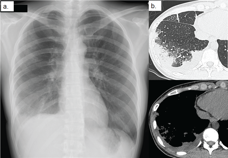
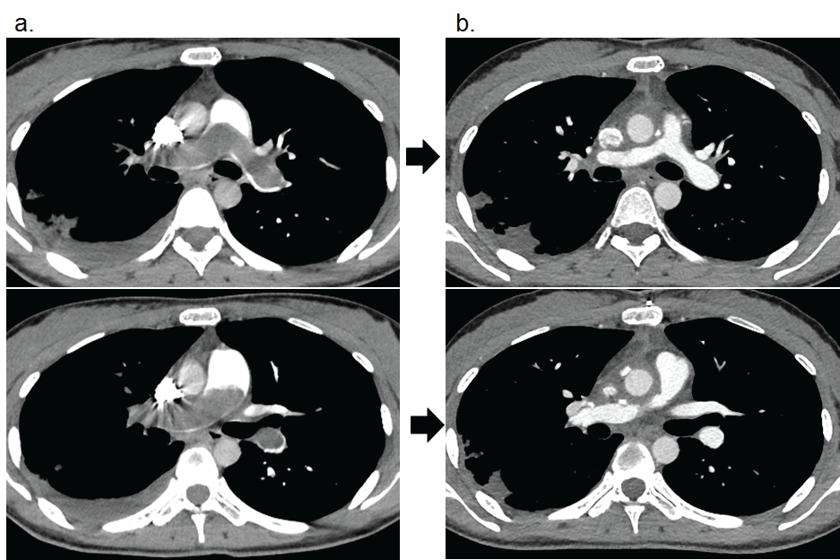
A 28-year-old woman presented to another hospital with fever, right chest pain, and cough without phlegm. Her chest pain was dull and was not exacerbated by deep respiration. She had a history of ovarian cyst removal and had a 0.5 pack/day smoking history for 5 years. Her chest X-ray showed right lower lobe consolidation, and she was treated with antibiotic therapy. However, her symptoms and chest X-ray did not improve, and she was referred to our hospital for a detailed investigation. The following clinical observations were recorded at initial examination: respiratory rate, 18 breaths/min; blood pressure, 104/54 mmHg; pulse rate 96/min; body temperature, 36.6 ℃; and oxygen saturation, 98%. Physical examination showed no abnormal findings except for decreased respiratory sounds in the right lower lung field on auscultation. Neither jugular venous distention nor pitting edema, which are characteristic of right ventricular heart failure, was noted. The laboratory data, including complete blood count, and blood biochemistry, showed no abnormalities. Only the C-reactive protein level was slightly high, and the D-dimer was significantly raised at 3.9 μg/ml. The results of an arterial blood gas analysis were as follows: PaCO2: 35.3 Torr; PaO2: 72.5 Torr. The serum human brain natriuretic peptide level was 1901 pg/mL. Her chest X-ray (Figure 1a) and non-contrast CT (Figure 1b) showed consolidation in the right lower lung field with pleural effusion. Decreased blood flow of the right lower lung field was not detected. A trans-bronchial biopsy was performed to aid in the diagnosis because there was a suspicion of organizing pneumonia, eosinophilic pneumonia, or other inflammatory pulmonary disease, but the results of bronchoalveolar lavage and the histological and cytological findings were not diagnostically useful. Next, the patient underwent an enhanced CT (Figure 2a) to evaluate the pleural disease. A large, saddle-filling defect extending into the right pulmonary artery and proximal lower lobe pulmonary artery branch was detected unexpectedly.
 Figure 1: a) Chest X-ray; b) Chest CT shows consolidation right lower lobe with pleural effusion. View Figure 1
 Figure 2: a) Enhanced chest CT before surgery, a large saddle filling defect extending into the right pulmonary artery as well as the proximal lower lobe pulmonary artery branch; b) After surgery, a large saddle filling defect was improved. View Figure 2
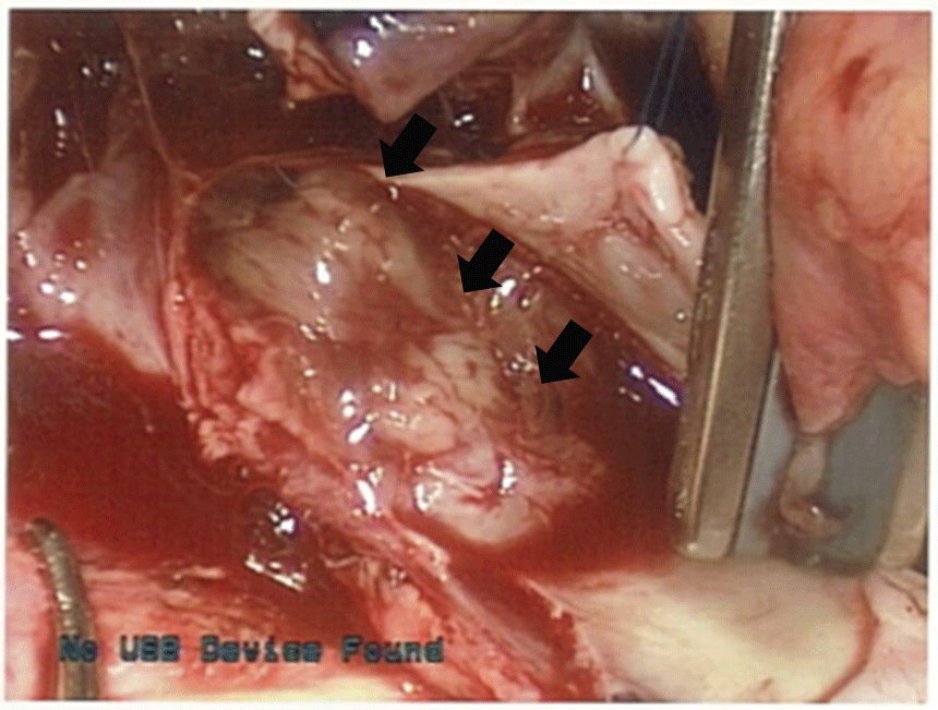
Although deep vein thrombosis in the legs was not detected, this large quantity of endoluminal material located proximally in the main pulmonary artery was considered to be a massive pulmonary artery thromboembolism; other diagnoses were not considered, and anticoagulation treatment was administered. Ultrasonic cardiogram (UCG) demonstrated mild tricuspid regurgitation, and the estimated pulmonary arterial pressure was 73 mmHg. The mean PA pressure and PA thrombus did not improve with anticoagulation therapy, and surgical intervention was performed. Surgery revealed that the main pulmonary artery was occupied by a myxomatous soft tissue tumor (Figure 3). The tumor was densely adherent to the pulmonary artery and was completely unresectable.
 Figure 3: Main pulmonary artery was occupied by myxomatous soft tissue tumor (arrow). View Figure 3
The microscopic findings showed atypical, spindle-shaped cells with a high mitotic activity, arranged as bundles and braids or irregularly (Figure 4). In addition, a myxoid and collagenized background was present. Immunohistochemical staining of the tumor cells was strongly positive for MDM2 and vimentin, but negative for desmin, smooth muscle actin, cytokeratin's (AE1/AE3), and CD34. According to these results, a diagnosis of pulmonary artery intimal sarcoma was made. After surgery, the radiographic findings, PA pressure, and chest pain improved (Figure 2b). Although we recommended chemotherapy or administration of a multi-targeted tyrosine kinase inhibitor, our patient did not agree to more aggressive therapy because there is no established chemotherapy or other aggressive treatment and she desired pregnancy. But 3 months later, pulmonary metastasis was detected, and she was treated with doxorubicin. Despite the 3 cycles of chemotherapy with doxorubicin, we could not control the disease activity and she passed away.
 Figure 4: Histological findings (hematoxylin eosin stain). a) x40; b) x200. Atypical spindle-shaped cells with high mitosis activity, arranged as bundles and braids or irregularly. View Figure 4
Pulmonary artery sarcomas are thought to be derived from endothelial cells of the pulmonary artery, and the term "intimal sarcoma" has been proposed recently. Pulmonary artery sarcomas are classified as undifferentiated, rhabdomyosarcoma, osteogenic sarcoma, angiosarcoma, fibrosarcoma, etc. The tumor's differentiation is classified according to the results of immunostaining. Most pulmonary artery intimal sarcomas arise from the pulmonary trunk and sometimes spread along the vessel's branches [1]. Pulmonary artery sarcomas are thought to be derived from endothelial cells of the pulmonary artery, and the term "intimal sarcoma" has been proposed recently. Pulmonary artery sarcomas are classified as undifferentiated, rhabdomyosarcoma, osteogenic sarcoma, angiosarcoma, fibrosarcoma, etc. The tumor's differentiation is classified according to the results of immunostaining. Most pulmonary artery intimal sarcomas arise from the pulmonary trunk and sometimes spread along the vessel's branches [1].
Huo reported 12 cases, and the mean age at diagnosis was 48.4 years [2]. He discussed the histological features. Two tumors were rhabdomyosarcoma, 4 were leiomyosarcoma, 1 was osteogenic sarcoma, 1 was angiosarcoma, and 4 were undifferentiated sarcoma. He reported that leiomyosarcoma had the longest survival, and rhabdomyosarcoma had the worst prognosis. Mussot reported 31 cases (16 male, mean age 56 years) and summarized the clinical characteristics [1]. The common symptoms were similar to those of pulmonary thromboembolism, including severe progressive or acute dyspnea (4/22), chest pain (4/31), fever (3/31), and syncope (1/31). Twenty-one patients had pulmonary hypertension, and 18 patients were suspected to have a pulmonary artery sarcoma.
In our case, making the diagnosis of pulmonary artery occlusion was very difficult, because the patient's symptoms were not severe, and she did not have poor oxygenation. At first, we diagnosed the patient with pneumonia with parapneumonic effusion, but this diagnosis became uncertain when antibiotic therapy was not effective. Therefore, we had to notice the laterality of pulmonary blood perfusion of the bilateral lung lower fields. Furthermore, the patient's inflammation markers were not particularly elevated, and thus, pulmonary artery embolism had to be considered. However, the patient was relatively young and had no risk factors such as obesity, pregnancy, or leg varices, and therefore, we had to consider that the cause of the pulmonary artery infarction was malignant vascular neoplasm. Parish, et al. reported 9 patients with pulmonary artery sarcomas whose presentation mimicked chronic thromboembolism [3]. They emphasized the efficacy of a combination of CT, MRI, and UCG to obtain a correct diagnosis. But in our case, tumor was only located intra-vascular area, so we could not diagnose as pulmonary artery sarcoma.
Based on previous reports, the histological type of our case was undifferentiated sarcoma, which is considered to have a poor prognosis [2]. Surgical resection was performed in 25 of the 31 patients, and 18 patients were administered adjuvant chemotherapy. Without aggressive therapy, the prognosis remains poor [1]. Blackmon discussed the outcome and determined that the mean survival was 36.5 ± 20.2 months after curative resection compared with 11 ± 3 months after incomplete resection [4]. The median survival was 24.7 ± 8.5 months with aggressive treatment compared with 8.0 ± 1.7 months with single-modality therapy. We attempted but were unable to perform a complete resection. Surgical resection is the only effective procedure, a few reports of the effectiveness of chemotherapy and radiotherapy are presented [4]. For patients with inoperable, metastatic or recurrent pulmonary artery sarcoma, chemotherapy with an anthracycline or its combinations with ifosfamide, gemcitabine, docetaxel and cisplatin are usually administered. Otherwise Funatsu reported the effectiveness of pazopanib for pulmonary artery sarcoma [5]. Our patient did not agree to more aggressive chemotherapy because she desired pregnancy, unfortunately multiple lung metastasis and regrowth of the surgical margin of right pulmonary artery were detected, the patient received three additional courses of chemotherapy with doxorubicin. Despite this chemotherapy, the tumors progressed, and the patient will be treated with other chemotherapeutic agents or multi-targeted tyrosine kinase inhibitor in the future.