

International Journal of Oral and Dental Health
Comparısıon Success Rate of Immedıate Implants at Fresh Extractıon Sockets and Conventıonal Implants
Elif Öncü*
Departmant of Perıodontolgy, Necmettin Erbakan Unıversity, Turkey
*Corresponding author:
Assist. Prof. Dr. Elif Öncü, PhD, DDS, Departmant of Perıodontolgy, Necmettin Erbakan Unıversity, Karaciğan Mah. Ankara Cad. No 74/A Karatay/Konya, Turkey, Tel: 0332 220 00 26, Fax: 0332 220 00 45, E-mail: oncu.elif@hotmail.com
Int J Oral Dent Health, IJODH-2-028, (Volume 2, Issue 2), Original Article; ISSN: 2469-5734
Received: March 20, 2016 | Accepted: May 27, 2016 | Published: May 29, 2016
Citation: Öncü E (2016) Comparısıon Success Rate of Immedıate Implants at Fresh Extractıon Sockets and Conventıonal Implants. Int J Oral Dent Health 2:028. 10.23937/2469-5734/1510028
Copyright: © 2016 Öncü E. This is an open-access article distributed under the terms of the Creative Commons Attribution License, which permits unrestricted use, distribution, and reproduction in any medium, provided the original author and source are credited.
Abstract
Objective: The aim of this presented study was to evaluate the survival and success rates implants with immediate and conventional placement. The second aim of this study was to evaluate and compared the crestal bone and gingival margin level around the implants in each groups.
Materials and methods: 30 immediate implants in 26 patients (test group) and 34 conventional implants in 26 patients (control group) were evaluated. All implants at least 12 months follow-up. The change in the level of crestal bone was measured on standardized digital panaromic radiographt and gingival margin recession values were evaluated in clinically, third months, and twelfth months for each patient. The measurements were statistically analyzed.
Results: Sixty-four implants healed without complication. Implants and restorations had a 100% survival rate during the study. Mean marginal bone resorption was calculated at 0.7 ± 0.5 mm for test group and 0.6 ± 0.6 for control group after at least 1 year in function. Both initially and after 12 months, gingival recession values were found similar for each group (p ≤ 0.05).
Conclusions: The results of this study demonstrated that the implant may be placed 2 mm below the crestal bone level to compensate the crestal bone loss. Immediat implant placement techniques are effective procedures as conventional implant placement.
Keywords
Immediate implant placement, Implants survivor rates, Gingival recession, Marginal bone level
Introduction
Immediate implant placement has gained popularity because it can reduce treatment time, number of surgeries and post-extraction bone loss [1,2]. Following tooth extraction, socket area shows series of physiologic processes, during the recovery time that passes between tooth extraction and the placement of the implant, the majority of the amount of bone resorption and gingival remodeling is confirmed, which is usually the cause of biological, aesthetic and functional damage [3]. Because of these factors, the technique for placement of implants immediately and early immediately after extraction was proposed as a way of maintaining the osseous complex of the surgical area [2,3]. Immediate implant placement refers to the placement of an implant into a tooth socket concurrently with the extraction [2-4]. Modeling of the alveolar ridge after extraction continues to occur following the implant placement [5,6].
The aim of this study was to evaluate the survival and success rates implants with immediate and conventional placement. The second aim of this study was to evaluate and compared the crestal bone and gingival margin level around the implants in each groups.
Materıals and Methods
Patient selection
The clinical and radiographical outcomes evaluated of 26 patients (16 male, 10 female) who treated at the department of Periodontology, School of Dentistry, University of Necmettin Erbakan Konya, Turkey with two or more adjacent or contralateral missing premolar and molar teeth were screened for eligibility to participate in this retrospective study. This retrospective study protocol was approved by the Ethics Committee of the Necmettin Erbakan University. 30 immediate implants in 26 patients (test group) and 34 conventional implants in 26 patients (control group) were evaluated. All implants at least 12 months follow-up. Additional surgery requirement, complications, advantages and disadvantages of immediate and conventional implant placement were compared. Patients record screened retrospectively who did not have any systemic health problem and did not need sinus floor augmentation, distraction osteogenesis and bone grafting had at least 2 adjacent premolar or molar teeth in the mandibula and maxilla. After extracted teeth and surgical preparation of sockets, implants placement immediatly in test group. The other implants were placed conventionaly in control group. All implants were evaluated in clinically and radiographically every twelveth months. All treatment steps were part of the routine procedures at the clinic, and no extra measures were made for this study.
Surgery procedure
Surgical procedures were performed by the same surgeon (E. Ö.). The surgical operations were carried out with local infiltration anesthesia (Ultracaine D-S®, Hoechst A.G, Turkey). After a crestal incision, a mucoperiosteal flap was elevated. Remaining teeth were carefully luxated with periotomes and teeth pulled out. Sockets were cleaned from granulation tissue and rinsed with saline. Implant sites were prepared in extraction sites, the implants were submerged 2 mm below the margins of the socket. The control groups implants were placed in the edentulous space as conventional. Mucoperiosteal flaps were brought to the original position, healing caps were not covered and flaps were sutured with 4/0 vicryl sutures. After the 7 days sutures were removed. Patients, the operation would lead to disruption in their daily lives in a subsequent period, and any discomfort or pain with the reported. They received their prescribed drugs regularly. Developed complications were’nt observed during the recovery period. Healing caps were placed in 3rd months.
Evaluation and criteria of success
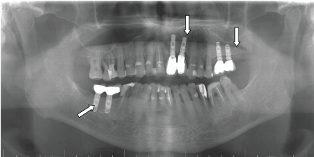
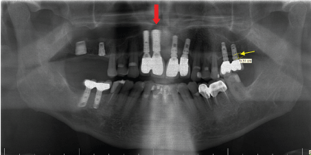
All implants were evaluated at 12 months after placement. During clinical recalls at 3 months, and 12 months changes in gingival margins were recorded with in millimeters by Williams Probe (Hu Friedy, Chicago, IL, USA) and bone loss evaluated in standart digital panaromic radiography (Figure 1). As part of the general routines, patients were enrolled in an individually designed maintenance care program for professional cleaning and examinations if needed. The change in the level of crestal marginal bone level was measured on standardized digital panaromic radiographt (Figure 2). All panoramic images were taken using the same panoramic machine (Eastman Kodak® 8000; Rochester, NY) by the same technician, according to the manufacturer’s reference guide. All images were recorded as JPEG files.
.
Figure 2: Red arrows shows that immediate implants, white arrows indicate the conventional implants. Yellow arrows indicate the measurement points.
View Figure 2
The upper corner of the coronal shoulder of the implant was used as reference point, and measurements from the reference point to the first bone contact of the implant were performed using a PC. An implant was accepted successful when (1) no sign of failure seemed in panoramic and periapical radiography, (2) no pain or symptoms of infection were present, (3) ≥ 2 mm bone loss, (4) gingival margin recession.
Statistical analysis
Statistical computations were carried out using IBM PASW/SPSS software (v.18.0.0 2009, IBM Corporation, Somers, NY, USA). Implants were included in the statistical analysis as independent values. Mean values and standard deviations were calculated for each variable and group. The difference between groups was analyzed with ANOVA and the difference within groups was analyzed with Student t-test.
Results
The study population consisted of 26 patients (16 male and 10 female) mean age 40.2 ± 11.5. Sixty-four implants healed without complication. Implants and restorations had a 100% survival rate during the study. The marginal bone level was positioned mean 0.5 ± 0.5 mm (range 0 to 2.1 mm) below the reference point at baseline for each group and 1.2 ± 0.6 mm after at least 1 year in function in test group and 1.1 ± 0.5 in control group. Mean marginal bone resorption was calculated at 0.7 ± 0.5 mm for test group and 0.6 ± 0.6 for control group after at least 1 year in function. Initially and ultimately measured values were not significantly different between groups (p ≤ 0.05) (Table 1).
![]()
Table 1: Statistical analysis between groups.
View Table 1
The mean gingival margin recession were 0.22 mm at the time of prothesis in control group and 0.20 mm in test group, 0.51 mm at 12 months in control group and 0.49 mm in test group. Both initially and after 12 months, gingival recession values were found similar for each group (p ≤ 0.05) (Table 1).
Dıscussıon
Immediate implant placement at fresh extraction sockets has become a common surgical protocol in clinical practice. This concept asserted reduced exposure of patients to surgery, limited physiological bone resorption and better gingival aesthetics outcomes [7,8]. Physiological dimensional changes occur in the alveolar ridge after tooth extraction and these changes usually occur within the first 3 months of socket healing [8-10]. These dimensional changes in the alveolar ridge may be prevented by placing immediate implant at at fresh extraction sockets [9-11].
Survival rates, reported in the literature as the avarege results of immediate implant protocols 2-year survival rate of 98.4%, 4-year implant-survival rate decreased to 97.5% in our study 1.5 year survival rate found 100% [12]. The dimensional changes occurring in alveolar crestal bone after tooth extraction and immediate implant placement have been evaluated in several clinical studies [8-18]. In a similar experimental study the vertical dimensional changes of the buccal bone wall were 2.1 ± 0.4 mm apical to the fixed landmark, after 12 weeks of healing and at the lingual wall, only minor changes were also observed [19]. Boticelli et al. have reported that 3.14 mm vertical bone resorption in their study after 4 months later after placing immediate implants [20,21]. In a smiller study, Blanco et al. 3 months later, they found 1.33 mm bone resorption [21]. In our study, the mean marginal bone resorption was calculated at 0.7 ± 0.5 mm for test group after at least 1 year in function. The results of this study was similar to other studies. In this retrospective study, the buccal and lingual areas were not separately evaluated. In this study all immediat implants were placed 2 mm below the crestal bone level. May be this method provided to compensate the crestal bone loss. Chen et al. evaluated the changes in the buccal gingiva recession retrospectively in immediately placed implant-supported restorations, with a mean follow-up of 18 months, reporting the occurrence of marginal tissue recession ( ≥ 1 mm) in one-third of the sites (33.3%) [22]. They reported that, the position of the implant shoulder in relation to the buccal bone plate was significantly associated with the occurrence of marginal recession. According to other similar study, gingival recessions were found 1.8 ± 0.83 mm in the buccally placed implants compared with only 0.6 ± 0.55 mm in those inserted lingually [10]. In this study it was found that the implants placed lingually. The mean gingival margin recession were found 0.51 mm in control group and 0.49 mm in test group at 18 months. This result similar with Kan et al. who reported that mean facial mucosal recession of 0.55 mm at the and of 1 year [23].
Conclusıon
Similar survival rates were observed for immediate and conventional placement, the present study indicated that immediat implant placement techniques are effective procedures as conventional implant placement. The implant may be placed 2 mm below the crestal bone level to compensate the crestal bone loss.
References
-
Schulte W, Heimke G (1976) The Tübinger immediate implant. Quintessenz 27: 17-23.
-
Esposito M, Grusovin MG, Polyzos IP, Felice P, Worthington HV (2010) Timing of implant placement after tooth extraction: immediate, immediate-delayed or delayed implants? A Cochrane systematic review. Eur J Oral Implantol 3: 189-205.
-
Qian J, Wennerberg A, Albrektsson T (2012) Reasons for marginal bone loss around oral implants. Clin Implant Dent Relat Res 14: 792-807.
-
Lang NP, Pun L, Lau KY, Li KY, Wong MC (2012) A systematic review on survival and success rates of implants placed immediately into fresh extraction sockets after at least 1 year. Clin Oral Implants Res 23 Suppl 5: 39-66.
-
Huber S, Rentsch-Kollàr A, Grogg F, Katsoulis J, Mericske R (2012) A 1-year controlled clinical trial of immediate implants placed in fresh extraction sockets: stability measurements and crestal bone level changes. Clin Implant Dent Relat Res 14: 491-500.
-
Meredith N (1998) Assessment of implant stability as a prognostic determinant. Int J Prosthodont 11: 491-501.
-
Becker W, Sennerby L, Bedrossian E, Becker BE, Lucchini JP (2005) Implant stability measurements for implants placed at the time of extraction: a cohort, prospective clinical trial. J Periodontol 76: 391-397.
-
Mijiritsky E, Mardinger O, Mazor Z, Chaushu G (2009) Immediate provisionalization of single-tooth implants in fresh-extraction sites at the maxillary esthetic zone: up to 6 years of follow-up. Implant Dent 18: 326-333.
-
Paolantonio M, Dolci M, Scarano A, d'Archivio D, di Placido G, et al. (2001) Immediate implantation in fresh extraction sockets. A controlled clinical and histological study in man. J Periodontol 72: 1560-1571.
-
Vignoletti F, Sanz M (2014) Immediate implants at fresh extraction sockets: from myth to reality. Periodontol 2000 66: 132-152.
-
Araújo MG, Lindhe J (2009) Ridge alterations following tooth extraction with and without flap elevation: an experimental study in the dog. Clin Oral Implants Res 20: 545-549.
-
Lang NP, Pun L, Lau KY, Li KY, Wong MC (2012) A systematic review on survival and success rates of implants placed immediately into fresh extraction sockets after at least 1 year. Clin Oral Implants Res 23 Suppl 5: 39-66.
-
Zix J, Kessler-Liechti G, Mericske-Stern R (2005) Stability measurements of 1-stage implants in the maxilla by means of resonance frequency analysis: a pilot study. Int J Oral Maxillofac Implants 20: 747-752.
-
Vidyasar L, Salms G, Apse P, Teibe U (2004) Dental Implant Stability at Stage I and II Surgery as Measured Using Resonance Frequency Analysis. Baltic dental and maxillofacial journal 6: 35-39.
-
Nedir R, Bischof M, Szmukler-Moncler S, Bernard JP, Samson J (2004) Predicting osseointegration by means of implant primary stability. Clin Oral Implants Res 15: 520-528.
-
Huang HM, Chiu CL, Yeh CY, Lee SY (2003) Factors influencing the resonance frequency of dental implants. J Oral Maxillofac Surg 61: 1184-1188.
-
Huang HM, Lee SY, Yeh CY, Lin CT (2002) Resonance frequency assessment of dental implant stability with various bone qualities: a numerical approach. Clin Oral Implants Res 13: 65-74.
-
Becker W, Sennerby L, Bedrossian E, Becker BE, Lucchini JP (2005) Implant stability measurements for implants placed at the time of extraction: a cohort, prospective clinical trial. J Periodontol 76: 391-397.
-
Araújo MG, Sukekava F, Wennström JL, Lindhe J (2006) Tissue modeling following implant placement in fresh extraction sockets. Clin Oral Implants Res 17: 615-624.
-
Botticelli D, Renzi A, Lindhe J, Berglundh T (2008) Implants in fresh extraction sockets: a prospective 5-year follow-up clinical study. Clin Oral Implants Res 19: 1226-1232.
-
Botticelli D, Persson LG, Lindhe J, Berglundh T (2006) Bone tissue formation adjacent to implants placed in fresh extraction sockets: an experimental study in dogs. Clin Oral Implants Res 17: 351-358.
-
Chen ST, Darby IB, Reynolds EC, Clement JG (2009) Immediate implant placement postextraction without flap elevation. J Periodontol 80: 163-172.
-
Kan JY, Roe P, Rungcharassaeng K, Patel RD, Waki T, ET AL. (2011) Classification of sagittal root position in relation to the anterior maxillary osseous housing for immediate implant placement: a cone beam computed tomography study. Int J Oral Maxillofac Implants 26: 873–876.
