

International Journal of Neurology and Neurotherapy
Lateral Basal Space and Interlaminar Fatty Compartment of the Deep Cervical Fascia in the Posterolateral Craniocervical Junction - An Anatomical Basis for the Surgery in the Lateral Skull Base
Katsuyoshi Shimizu1*, Akira Wada1, Mika Kushamae1, Ryo Irie1, Yuta Kawauchi1, Yu Kato1,2, Kazuki Iizuka1, Minako Kubo1, Yu Sakamoto1, Hiromitsu Ezure2, Naruhito Otsuka2, Tohru Mizutani1
1Department of Neurosurgery, Showa University School of Medicine, Japan
2Department of Anatomy, Showa University School of Medicine, Japan
*Corresponding author:
Katsuyoshi Shimizu, Department of Neurosurgery, Showa University School of Medicine, 5-8 Hatanodai 1, Shinagawa-ku, Tokyo 142-8666, Japan, E-mail: katsuyoshis@aol.com
Int J Neurol Neurother, IJNN-3-038, (Volume 3, Issue 1), Research Article; ISSN: 2378-3001
Received: January 05, 2016 | Accepted: January 25, 2016 | Published: January 27, 2016
Citation: Shimizu K, Wada A, Kushamae M, Irie R, Kawauchi Y, et al. (2016) Lateral Basal Space and Interlaminar Fatty Compartment of the Deep Cervical Fascia in the Posterolateral Craniocervical Junction - An Anatomical Basis for the Surgery in the Lateral Skull Base. Int J Neurol Neurother 3:038. 10.23937/2378-3001/3/1/1038
Copyright: © 2016 Shimizu K, et al. This is an open-access article distributed under the terms of the Creative Commons Attribution License, which permits unrestricted use, distribution, and reproduction in any medium, provided the original author and source are credited.
Abstract
In the lateral suboccipital area, the restricted space filled with thick muscles, and dangerous structures prevents neurosurgeons to attain safe and successful surgery. Now we have focus on the disposition of fascial layers and potential spaces of the deep cervical fascia in the posterolateral region of the craniocervical junction, which has barely been described before. Investigation with 32 lateral suboccipital surgical cases and 7 cadaveric dissections has revealed the detailed anatomy in this area. We have also identified the interlaminar fatty compartment expanding between the skull base and the carotid sheath, which was partially described in previous reports. In addition, we have recognized the lateral basal space of the deep cervical fascia which is occupied by a part of this fatty connective tissue. Proper preparation of the fatty compartment and adjacent fascial layers enables us to take advantage of this lateral basal space widely opened for approaching to the lateral base of the cranium. We believe that the concept of this space contribute to the advance of the surgery in the posterolateral craniocervical junction.
Keywords
Craniocervical junction, Deep cervical fascia, Lateral basal approach, Skull base surgery
Abbreviations
C1: first cervical vertebra; DCF: Deep Cervical Fascia; ILFD: Interlaminar Fatty Compartment of the Deep cervical fascia; LBSD: Lateral Basal Space of the Deep cervical fascia; LOC: Longissimus Capitis; OA: Occipital Artery; pMD: posterior venter of the digastric muscle; SCM: Sternocleidomastoid; SPC: Splenius Capitis; SPO: superior oblique muscle; SSC: Semispinalis Capitis
Introduction
For the surgery on the posterolateral craniocervical region, various kinds of lateral suboccipital, retrosigmoid approaches are attempted such as far lateral, transcondylar, transcondylar fossa approaches and so on [1]. Additionally, harvesting the occipital artery (OA) as an important doner artery for extracranial - intracranial bypass surgery, a superficial approach to this area is now common technique for neurosurgeons [2]. To successfully reach the posterolateral area of the craniocervical junction, the accurate knowledge about the anatomical configuration of the deep cervical fascia (DCF) and surrounding important structures is essential. Those of the anterior craniocervical junction are well described in numerous previous literatures and text books because infections and cancer cells ascend along the fascial planes to the skull base [3-5]. However, only few articles have contributed the anatomical information concerning the posterior craniocervical junction [6,7]. The detailed anatomy of the DCF and related potential spaces in this transitional area remains to be revealed.
Here, we describe in particular the disposition of the DCF in the posterolateral part of the craniocervical junction demonstrating a new concept of the interlaminar fatty compartment and the lateral basal space of the DCF (ILFD, and LBSD) studied by the operative cases and human adult cadavers. This small fatty space is of great importance for the safe and successful approach to the lateral skull base and the secure preservation of the OA
Materials and Methods
All patients consented to have their clinical data subsequently submitted to a journal. Surgical treatments with the lateral suboccipital approaches were recently performed on 32 adult patients. Intraoperative findings and postoperative video observations were adopted in all patients (20 male and 12 female; age rage, 34-82 years). Twenty-nine cases underwent the lateral basal approach for microvascular decompression on hemifacial spasms and trigeminal neuralgias. Three cases were treated by the lateral suboccipital retrosigmoid approach for vestibular schwannomas and a cerebellopontine angle meningioma. The surgical procedures were carefully advanced separating each fascial plane to recognize the proper layers and spaces. Seven adult cadavers (4 male and 3 female) were also used, which were donated to Showa University School of Medicine for education and research.
Results
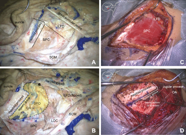
In the operative case, appropriate skin incision was performed on the lateral suboccipital region and the superficial layer of the DCF was disclosed. This layer, including the sternocleidomastoid muscle (SCM) was sharply dissected from the mastoid process and the superior nuchal line at its attachment. Subsequently, the splenius capitis (SPC) was exposed underneath embedded in the lateral part of the outmost layer of the prevertebral layer of the DCF. It also contained the longissimus capitis (LOC) muscle immediately under the SPC. At the same time, we necessarily recognized the fatty tissue abutting on the anterior margin of the SPC (Figure 1C). This compartment could be seen in all cases, which was composed of thick and fatty connective tissue. It was located between the superficial and the prevertebral layer of the DCF sneaking under the SPC and the LOC. Superficially, it seemed to descend anteriorly to the fatty connective tissue surrounding the carotid sheath under the SCM. Deeply it was obviously bound by the thick fascial connective tissue over the superior oblique muscle (SPO) attached to the transverse process of the first cervical vertebra (C1). Thus this fatty compartment intervenes between the rear side of mastoid tip and the transverse process of C1 on the bottom of the cranium (Figure 2). It also enclosed the posterior venter of the digastric muscle (pMD) at its attachment to the digastric groove fusing to the periosteum at the bottom of the mastoid process. We termed this compartment as the interlaminar fatty compartment of the DCF (ILFD).
.
Figure 1: (A, B) Pictures indicating the ILFD and LBSD in the right posterolateral craniocervical junction demonstrated in the cadaver dissection; (C, D) the lateral basal approach for microvascular decompression on hemifacial spasm. A and C, The SCM separation from mastoid process discloses the ILFD is located besides the SPC.; B, The SPC, LOC and the ILFD are dissected to identify the LBSD (yellow area margined by dotted line). The pDM is removed. The black dot is the transverse process of C1; D, Of note is the widely disclosed lateral bottom of cranium in the separated LBSD even through a small skin incision (8,9). The pDM is removed.
View Figure 1
.
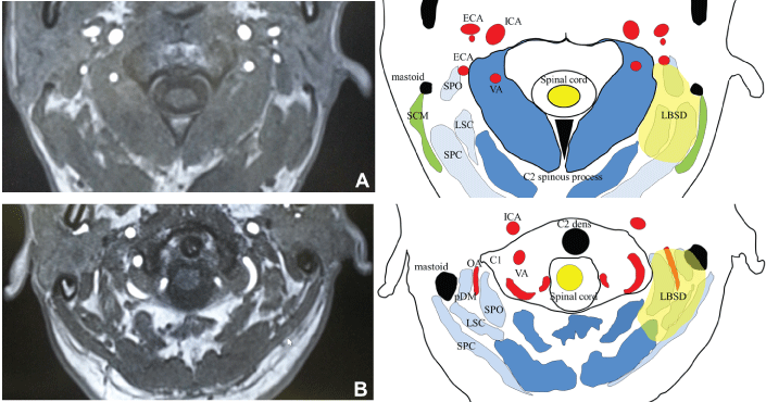
Figure 2: Illustrations depicting the disposition of the fascial layers and potential spaces of the deep cervical fascia with comparable axial MR TOF images. The circled area is the LBSD originally filled with the ILFD, which is expanded up to the yellow area after cutting down the SPC, LOC, and the ILFD. (A) section at the level of the second cervical vertebra; (B) section at the level between skull base and C1.
View Figure 2
The occipital artery (OA) was always secured in this compartment emerging from under the pDM (Figure 1D and Figure 2B). Then the SPC and the LOC were cut down from the mastoid process. Subsequently adequate dissection of this fatty compartment provided the wide and safe space which is valuable for approaching to the lateral bottom of the cranium even through the restricted corridor adopted for the skull base surgery [8,9] (Fgure 2). We also termed this space originally occupied by the ILFD as the lateral basal space of the DCF (LBSD). For the optimal craniotomy for the lateral basal approach, the partial dissection of the SPO and its thick fascia was sometimes performed safely at their attachment to the transverse process us of C1 [9].
The cadaver dissections widely spread the sight more anteriorly than that obtained from operative cases by cutting down the SCM and pDM (Figure 1A and Figure 1B). The ILFD was identified to occupy the LBSD and to expand superficially to the fatty connective tissue on the lateral surface of the carotid sheath, and deeply to the posterior part of the carotid sheath enveloping the OAin all cases. Simultaneously, the LBSD was revealed to be demarcated superiorly by the lateral bottom of the occipital cranium, laterally by the SCM and medially by the SPO attaching to the transverse process of C1 covered by the alar fascia.
Discussion
Deep cervical fascia
There is no generally accepted definition on the semantics of fasciae, layers, or spaces. Interdisciplinary differences in the foci of this area make it more confused since the classical description in 1938 [4,10]. Although the anatomy of the DCF and related potential spaces has been controversial, the widely believed concept is as follows [4].
The superficial layer of the deep cervical fascia contains the trapezius and the SCM. Deep inside this layer, other layers form two compartments. Anteriorly, the pretracheal layer forms the visceral compartment containing the trachea, esophagus, thyroid gland, and adjacent structures. The pretracheal fascia has apparently been used synonymously with visceral fascia. Posteriorly, the prevertebral layer encloses the vertebral column and its associated muscles. Between these two structures, the condensation of the connective tissues around the carotid artery, jugular vein, and vagus nerve forms the carotid sheathon each side. The outmost prevertebral layer of the DCF is generally regarded to enclose the semispinalis capitis (SSC) and the SPC [10].
In all clinical cases of the present study, the LOC has been simultaneously separated with the SPC. Thus, we have suggested that the LOC was also enveloped by the prevertebral layer of the DCF. The prevertebral layer of the DCF has been reported to attach medially to the external occipital protuberance and crest, and laterally to the mastoid process [10]. In this study, the lateral attachment has been described more precisely as the lateral part of the inferior nuchal line, which continues basally along the digastric groove to the level of the lateral edge of the jugular process of the skull base. Under the SCM, the SPC and the ILFC have been situated side by side seeming to be embedded together in the prevertebral layer of the DCF. The ILFC has also been closely connected to the thick fascial lamina of the SPO in the prevertebral layer of the DCF attaching to the basal surface of the mastoid. Thus, it is difficult to discern whether the ILFC could be a part of the prevertebral layer of the DCF demarcated by the alar fascia or a fatty compartment between the superficial and prevertebral layers. This limitation, in dissecting out the fascial layer, has been indicated on the cadaver dissection even under the operative microscope [6,10]. We have also found the same difficulties even in alive operative cases. On the other hand, the pDM obviously has its own particular fascia and is enclosed in the ILFC along the digastric groove.
Carotid sheath
Grodinsky and Holyoke suggested that the superficial layer of the DCF passes medially splitting into the two laminae deep to the SCM forming anterolateral and posterior walls of the carotid sheath, which finally merge to the pretracheal layer of the DCF as the medial wall of the carotid sheath [3,4]. Recently, advanced radiologic and cadaveric studies have described that the carotid sheath is composed of all three layers of the DCF; the superficial, pretracheal, and prevertebral layers [6,7]. It is not surprising because dispositions and fusions of fasciae form fascial spaces, which sometimes is barely distinguished from areas of relatively loose connective tissue. The alar fascia, defined as a loose areolar connective tissue within the retropharyngeal region, could divide the deep space into some particular compartments between the visceral and the prevertebral fascia, which are readily identified with the advances in the radiological and surgical technology [7,10]. We have revealed the fatty compartment adjacent to the anterior margin of the SPC, which descends superficially into the surrounding fatty tissue and deeply fuses with the posterolateral part of the carotid sheath. These findings are consistent with the recent reports concerning this area [6,7,10].
The lateral basal space of the deep cervical fascia (LBSD) and the interlaminar fatty compartment of the deep cervical fascia (ILFD)
Many spaces are closely described in the peripharyneal area deep under the superficial DCF [3,4,10]. Although the organization of connective tissues in the anteromedial region of the craniocervical region has been well documented in the literatures [5,10], those of the posterolateral one has barely been reported in detail [6,7]. The alar fascia is generally noticed to be a division of the deep layer of the DCF spanning between the transverse processes of the cervical vertebrae and fusing laterally with the carotid sheath [7,10]. However, it still remains inconsistent whether the alar fascia extends to the skull base [6,7]. Recent study has indicated that the alar fascia begins at the level of C1 whereas loose fibroareolar connective tissue occupies the space between the inferior nuchal line and the skull base [7]. We have demonstrated the details about the fascial attachment of DCF at the posterolateral base of the skull. A tight fascial attachment has been identified on the lateral part of the inferior nuchal line descending the digastric groove at the bottom of the mastoid process to the lateral edge of the jugular process. Medially, there has been found no tight connection in the skull base, which is compatible with the aforementioned report [7].
The LBSD is the space widely opened by cutting down the ILFD, SPO and LOC immediately behind the mastoid tip. It is demarcated laterally by the SCM, medially by the fascial layer over the SPO, and is also roofed by the bottom of the cranium. Thus, the LBSD faces to the bony surface which is necessarily exposed for the maximum operative field for the lateral suboccipital craniotomies. After preserving the OA, we can take advantage of this space free from injuring dangerous structures, because the vertebral artery on the lamina of C1 arch is also covered by the medial wall of the thick muscular layer of SPO. Manipulation of the condyle emissary vein is not necessary either for approaching the lateral skull base by utilizing the LBSD lateral to the condyle fossa. The ILFD originally occupies the LBSD and expands back and forth merging to some other fatty spaces previously demonstrated. Grodinsky and Holylke previously localized the distinct space between the superficial and prevertebral layers of the DCF in the posterior triangle [3,4]. However it was described mainly below the hyoid bone. Recently, Zhang et al. have demonstrated the two potential spaces in the suboccipital region from the superior nuchal line to the second cervical vertebra [6]. One is the space termed the suboccipital space which runs along the posterior midline and the other is a paired space between the prevertebral DCF and the SCM. The latter one is practically the upper extension of the space shown by Grodinsky and Holylke. This space, occupied by the fatty tissue, connects anteromedially to the carotid sheath, and posterolaterally to the fatty space between the SCM and the trapezius. It is situated inferior to the original LBSD blending with the fatty compartment in the lower cervical portion [6]. We also demonstrated that the OA is embedded deep in the ILFC. In all surgical cases, the transverse portion of the OA is secured emerging from under the pDM. Fukuda et al. demonstrated that the OA ran in a single fatty connective tissue layer between the SPC and the SSC [2]. They also implied that this thin fatty layer was a posterior extension of the styloid diaphragm, which was termed by Bejjani et al. [11]. The styloid diaphragm was defined as the space in the infratemporal fossa bordered by the pDM, the styloid muscle group and the SCM, which represented the area anterior to the carotid sheath. Apparently, the OA runs through the layer between the SPC and SCC after passing the ILFC occupying the LBSD. Consequently, we have suggested the LBSD is intervening between the two spaces previously termed styloid diaphragm and its thin posterior extension [2,11].
Conclusions
Herein, we have devotedly focused on the area closely adjacent to the lateral bottom of the cranium identifying the anatomical features considerable for the skull base surgery. Until now, there has never been a universal concept concerning the fascial spaces or layers in the DCF in this area. Therefore, the description in this article is possibly prone to be arbitrary one. Nevertheless, we have dared to suggest new concepts of the LBSD and the ILFC closely adjacent to the posterolateral bottom of the cranium. Identifying these compartments will not only improve surgical management in this area, but will also provide anatomical basis for surgeons to design approaches to the lesions in the posterolateral craniocervical junction.
References
-
Matsushima T, Kawashima M, Masuoka J, Mineta T, Inoue T (2010) Transcondylar fossa (supracondylar transjugular tubercle) approach: anatomic basis for the approach, surgical procedures, and surgical experience. Skull Base 20: 83-91.
-
Fukuda H, Evins AI, Burrell JC, Stieg PE, Bernardo A (2014) A safe and effective technique for harvesting the occipital artery for posterior fossa bypass surgery: A cadaveric study. World Neurosurgery 82(3-4): e459-e465.
-
Grodinsky M, Holyoke E (1983) The fasciae and fascial spaces of the head, neck and adjacent regions. Am J Anat 63: 367-408.
-
Hollinshead WH (1982) Anatomy for surgeons: Vol 1 The head and neck, 3rd ed. Harper & Row, Publishers, Philadelphia Chapter5: 269-289.
-
Nash L, Nicholson HD, Zhang M (2005) Does the investing layer of the deep cervical fascia exist? Anesthesiology 103: 962-968.
-
Zhang M, Lee AS (2002) The investing layer of the deep cervical fascia does not exist between the sternocleidomastoid and trapezius muscles. Otolaryngol Head Neck Surg 127: 452-454.
-
Scali F, Nash LG, Pontell ME (2015) Defining the Morphology and Distribution of the Alar Fascia: A Sheet Plastination Investigation. Ann Otol Rhinol Laryngol 124: 814-819.
-
Shimizu K, Matsumoto M, Wada A, Sugiyama T, Tanioka D, et al. (2015) Supine No-Retractor Method in Microvascular Decompression for Hemifacial Spasm: Results of 100 Consecutive Operations. J Neurol Surg B Skull Base 76: 202-207.
-
Shimizu K, Matsumoto M, Wada A, Mizutani T (2015) Lateral basal approach with a supine, no-retractor method for microvascular decompression for hemifacial spasm. Acta Neurochir (Wien) 157: 803-806.
-
Guidera AK, Dawes PJ, Fong A, Stringer MD (2014) Head and neck fascia and compartments: no space for spaces. Head Neck 36: 1058-1068.
-
Bejjani GK, Sullivan B, Salas-Lopez E, Abello J, Wright DC, et al. (1998) Surgical anatomy of the infratemporal fossa: the styloid diaphragm revisited. Neurosurgery 43: 842-852.
