A 70-year-old gentleman with a background of ischaemic heart disease and esophagectomy was admitted to Critical Care Unit with hypoxemic respiratory failure secondary to COVID pneumonitis. Prior to Critical Care admission, he was discovered to have deep venous thrombosis and he was commenced on treatment dose low molecular weight heparin.
The patient was hemodynamically stable and required high flow oxygen therapy for 72 hours. He had a sudden hemodynamic collapse whilst receiving high flow oxygen therapy. An ICU registrar performed a bedside echocardiography (Figure 1) that revealed a large volume blood clot occupying the Right Ventricular (RV) inlet. There was also ECHO evidence of Dilated RV; Dilated Right Atrium and RV pressure overload with a completely obliterated LV cavity.
This case supports the emerging evidence of increased risk of thrombosis in COVID pneumonitis. The benefit of therapeutic anticoagulation in these patients is not entirely clear and remains a research question. This case also highlights the importance of basic echo skills in critical care as this scan was performed by an amateur echocardiographer.
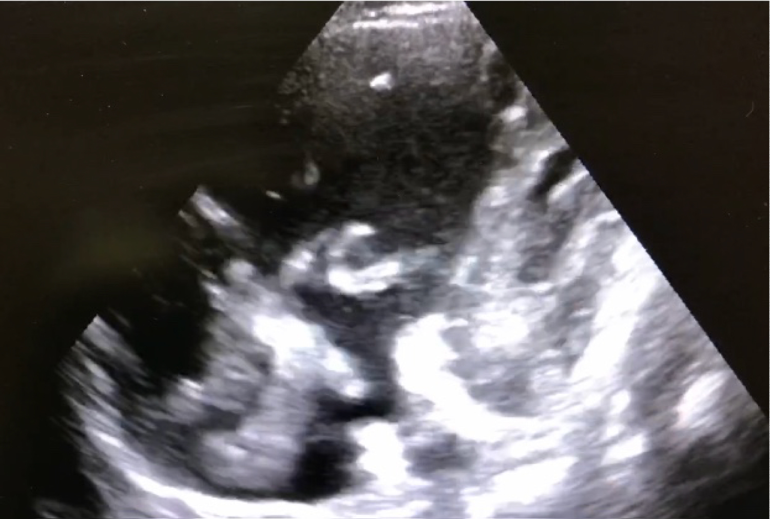
Figure 1: Echocardiography.